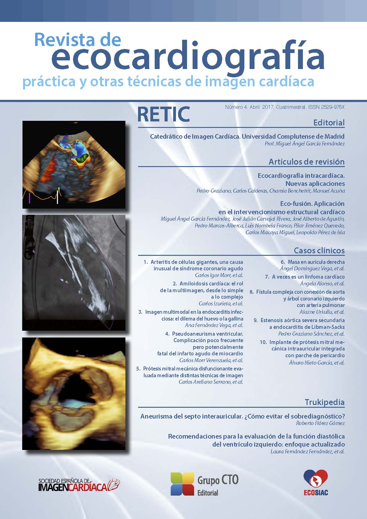
Eco-fusión. Aplicación en el intervencionismo estructural cardíaco
DOI:
https://doi.org/10.37615/retic.n4a3Palabras clave:
imágenes de fusión, enfermedades estructurales del corazón, guía intervencionista.Resumen
La evolución en el intervencionismo estructural percutáneo ha generado un desarrollo paralelo en las técnicas de imagen avanzada. La ecocardiografía en el intervencionismo estructural juega un papel básico en la selección de los pacientes, en la valoración durante el procedimiento y en el análisis inmediato de los resultados y la detección precoz de complicaciones. Las imágenes de fusión eco/fluoroscopia aparecen como una herramienta complementaria en la que dos técnicas con imágenes dinámicas se complementan en una sola imagen con el fin de orientar, disminuir el tiempo de intervención y disminuir las complicaciones.
Descargas
Métricas
Citas
Clegg S, Salcedo E, Quaife R, Carrol J. Imaging in structural heart disease. En: Lasala J, Rogers J. Interventional procedures for adult and structural heart disease. 1.ª ed. Elsevier-Saunders, 2014; 7-28.
Carminati M, Agnifili M, Arcidiacono C, et al. Role of imaging in interventions on structural heart disease. Expert Review of Cardiovascular Therapy 2013; 11: 1659-1676. DOI: https://doi.org/10.1586/14779072.2013.854166
Perk G, Lang RM, García-Fernández MA, et al. Use of real time three-dimensional transesophageal echocardiography in intracardiac catheter based interventions. J Am Soc Echocardiogr 2009; 22 (8): 865-882. DOI: https://doi.org/10.1016/j.echo.2009.04.031
Thaden J, Sanon S, Geske J, et al. Echocardiographic and fluoroscopic fusion imaging for procedural guidance: An overview and early clinical experience. Journal of American Society of Echocardiography 2016; 29: 503-512. DOI: https://doi.org/10.1016/j.echo.2016.01.013
Gaemperli O, Schepis T, Kalff V, et al. Validation of a new cardiac image fusion software for three-dimensional integration of myocardial perfusion SPECT and stand-alone 64-slice CT angiography. European Journal of Nuclear and Medicine Molecular Imaging 2007; 34: 1097-1106. DOI: https://doi.org/10.1007/s00259-006-0342-9
White JA, Fine N, Gula LJ, et al. Fused whole-heart coronary and myocardial scar imaging using 3-TCMR. Implications for planning of cardiac resynchronization therapy and coronary revascularization. JACC Cardiovascular Imaging 2010; 3: 921-930. DOI: https://doi.org/10.1016/j.jcmg.2010.05.014
Tanis W, Scholtens A, Habets J, et al. CT angiography and (1)(8)FFDG-PET fusion imaging for prosthetic heart valve endocarditis. JACC Cardiovascular Imaging 2013; 6: 1008-1013. DOI: https://doi.org/10.1016/j.jcmg.2013.07.004
Arujuna A, Housden R, Ma Y, et al. Novel system for real-time integration of 3D-Echocardiography and fluoroscopy for image guided cardiac interventions: preclinical Validation and clinical feasibility evaluation. IEEE Journal of traslational engineering in health and medicine 2014; 2: 110. DOI: https://doi.org/10.1109/JTEHM.2014.2303799
Balzer J, Zeus T, Hellhammer K, et al. Initial clinical experience using the Echonavigator® – system during structural heart disease interventions. World Journal of Cardiology2015; 7 (9): 562-570. DOI: https://doi.org/10.4330/wjc.v7.i9.562
Feldman T, Hellig F, Mollman H. Structural heart interventions: the state of the art and beyond. Eurointervention 2016; 12: X6. DOI: https://doi.org/10.4244/EIJV12SXA1
Palacios I, Arzamendi D. Intervencionismo en cardiopatía estructural. Más allá de la terapia valvular transcateter. Revista española de cardiología 2012; 65 (5): 405-413. DOI: https://doi.org/10.1016/j.recesp.2011.12.022
Afzal S, Veulemans V, Balzer J, et al. Safety and efficacy of trans-septal puncture guided by real-time fusion of echocardiography and fluoroscopy. Netherland Heart Journal. Neth Heart J 2017; 25 (2): 131-136. DOI: https://doi.org/10.1007/s12471-016-0937-0
Vahanian A, Alfieri O, Andretti F, et al, ESC Committee for Practice Guidelines (CPG); Joint Task Force on the Management of Valvular Heart Disease of the European Society of Cardiology (ESC); European Association for Cardio-Thoracic Surgery (EACTS). Guidelines on the management of valvular heart disease: the Joint Task Force on the Management of Valvular Heart Disease of the European Society of Cardiology (ESC) and the European Association for Cardio-Thoracic Surgery (EACTS). European Journal of Car- diothoracic Surgery 2012; 42: S1-44.
Sundermann S, Biaggi P, Grunenfelder J, et al. Safety and feasibility of novel technology fusing echocardiography and fluoroscopy images during Mitraclip interventions. Eurointervention 2014; 9: 1210-1216. DOI: https://doi.org/10.4244/EIJV9I10A203
García-Fernández MA, Cortés M, García-Robles JA, et al. Utility of real-time three-dimensional transesophageal echocardiography in evaluating the success of percutaneous transcatheter closure of mitral paravalvular leaks. J Am Soc Echocardiogr 2010; 23 (1): 26-32. DOI: https://doi.org/10.1016/j.echo.2009.09.028
Franco E, Almería C, de Agustín JA, Arreo Del Val V, Gómez de Diego JJ, García Fernández MÁ, Macaya C, Pérez de Isla L, García E. Three-Dimension al Color Doppler Transesophageal Echocardiography for Mitral Paravalvular Leak Quantification and Evaluation of Percutaneous Closure Success Jour- nal of American Society of Echocardiography 2014; 27 (11): 1153-1163. DOI: https://doi.org/10.1016/j.echo.2014.08.019
Cortés M, García E, García-Fernandez MA, et al. Usefulness of transesophageal echocardiography in percutaneous transcatheter repairs of paravalvular mitral regurgitation. American Journal of Cardiology 2008; 101 (3): 382-386. DOI: https://doi.org/10.1016/j.amjcard.2007.08.052
Nucifora G, Faletra FF, Regoli F, et al. Evaluation of the left atrial appendage with realtime 3-dimensional transesophageal echocardiography: implications for catheter-based left atrial appendage closure. Circulation Cardiovas- cular Imaging 2011; 4: 514-523. DOI: https://doi.org/10.1161/CIRCIMAGING.111.963892
Gafoor S, Schulz P, Heuer L, et al. Use of Echo-Navigator, a novel echocardiography-fluoroscopy overlay system, for transseptal puncture and left atrial appendage occlusion. Journal of Interventional Cardiology 2015; 28: 215-217. DOI: https://doi.org/10.1111/joic.12170
Jungen C, Zeus T, Balzer J, et al. Left atrial appendage closure guided by integrated echocardiography and fluoroscopy imaging reduces radiation exposure. Plos One 2015; 10: 1-13. DOI: https://doi.org/10.1371/journal.pone.0140386
García-Fernández M, De Agustín A, Pérez de Isla L. Eco-Xray fusion in left atrial appendage closure. Revista Española de Cardiología 2016. Article in press. DOI: https://doi.org/10.1016/j.rec.2016.05.020
Kempfert J, Noettling A, John M, et al. Automatically segmented Dyna-CT: enhanced imaging during transcatheter aortic valve implantation. Journal of American Collegue of Cardiology 2011; 58: e211. DOI: https://doi.org/10.1016/j.jacc.2011.05.065
Balzer J, Hall S, Rassaf T, et al. Feasibility, safety and efficacy of real-time three dimensional transoesophageal echocardiography for guiding device closure of interatrial communications: Initial clinical experience and impact on radiation exposure. European Journal of echocardiography 2010; 11: 1-8. DOI: https://doi.org/10.1093/ejechocard/jep116
Jone P, Ross M, Bracken J, et al. Feasibility and safety of using a fused echocardiography/Fluoroscopy imaging system in patients with congenital heart disease. Journal of American society of echocardiography 2016; 5: 13-21. DOI: https://doi.org/10.1016/j.echo.2016.03.014
Faletra FF, Pedrazzini G, Pasotti E, et al. Echocardiography-X-Ray Image Fu- sion. JACC Cardiovascular imaging 2016; 9 (9): 1114-1117. DOI: https://doi.org/10.1016/j.jcmg.2015.09.022
Avenatti E, Barker C, Little S. Tricuspid regurgitation repair with a Mitraclip device: transoesophageal echocardiography. Eur Heart J Cardiovasc Imaging 2017. Epub a head of print.
Muller D, Farivar R, Jansz P, et al. Transcatheter mitral valve replacement for patients with symptomatic mitral regurgitation. JACC 2017; 69: 381- 391. DOI: https://doi.org/10.1016/j.jacc.2016.10.068
Descargas
Publicado
Cómo citar
Número
Sección
Licencia
Derechos de autor 2017 Miguel Ángel García Fernández , José Julián Carvajal Rivera, José Alberto de Agustín , Pedro Marcos-Alberca , Luis Nombela Franco , Pilar Jiménez Quevedo , Carlos Macaya Miguel , Leopoldo Pérez de Isla

Esta obra está bajo una licencia internacional Creative Commons Atribución-NoComercial-SinDerivadas 4.0.
RETIC se distribuye bajo la licencia Creative Commons Reconocimiento-NoComercial-SinDerivadas 4.0 Internacional (CC BY-NC-ND 4.0) https://creativecommons.org/licenses/by-nc-nd/4.0 que permite compartir, copiar y redistribuir el material en cualquier medio o formato, bajo los siguientes términos:
- Reconocimiento: debe otorgar el crédito correspondiente, proporcionar un enlace a la licencia e indicar si se realizaron cambios. Puede hacerlo de cualquier manera razonable, pero no de ninguna manera que sugiera que el licenciante lo respalda a usted o su uso.
- No comercial: no puede utilizar el material con fines comerciales.
- No Derivados: si remezcla, transforma o construye sobre el material, no puede distribuir el material modificado.
- Sin restricciones adicionales: no puede aplicar términos legales o medidas tecnológicas que restrinjan legalmente a otros de hacer cualquier cosa que permita la licencia.


