
Echocardiography in the assesment of multiple papillary fibroelastoma
DOI:
https://doi.org/10.37615/retic.v6n3a6Keywords:
multiple papillary fibroelastoma, echocardiogram, aortic valveAbstract
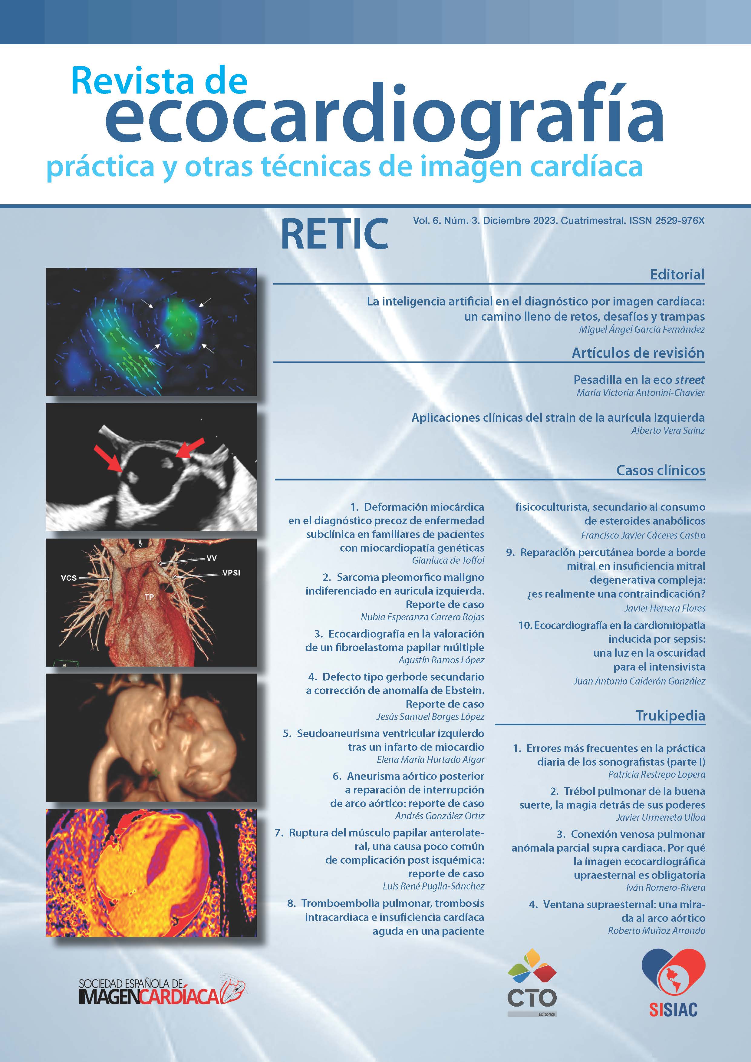
Fibroelastoma is the second most common primary benign cardiac tumor. It can be multiple; the aortic valve is the most frequently affected location and the most important complication is systemic embolism. We present the case of a 67-year-old woman who attended the emergency room due to heart failure. In the study, the echocardiogram showed a nodular small mass in the left coronary leaflet of the aortic valve suggestive of fibroelastoma. The transesophageal echocardiogram confirmed the finding and found a second mass in the non-coronary leaflet, for which to the final diagnosis of multiple papillary fibroelastoma.
Downloads
Metrics
References
Mariscalco G, Bruno VD, Borsani P, Dominici C, Sala A. Papillary fibroelastoma: insight to a primary cardiac valve tumor. J Card Surg. 2010;25(2):198-205. https://doi.org/10.1111/j.1540-8191.2009.00993.x DOI: https://doi.org/10.1111/j.1540-8191.2009.00993.x
Tsugu T, Nagatomo Y, Endo J, et al. Multiple papillary fibroelastomas attached to left ventricular side and aortic side of the aortic valve: A report of new case and literature review. Echocardiography. 2019;36(6):1194-1199. https://doi.org/10.1111/echo.14350 DOI: https://doi.org/10.1111/echo.14350
Tyebally S, Chen D, Bhattacharyya S, et al. Cardiac Tumors: JACC CardioOncology State-of-the-Art Review. JACC CardioOncol. 2020;2(2):293-311. Published 2020 Jun 16. https://doi.org/10.1016/j.jaccao.2020.05.009 DOI: https://doi.org/10.1016/j.jaccao.2020.05.009
Jahnke C, Hamdan A, Fleck E, Paetsch I. Tissue characterization of a suspected aortic valve fibroelastoma with cardiac magnetic resonance imaging. Circ Cardiovasc Imaging. 2008;1(1):87-88. https://doi.org/10.1161/CIRCIMAGING.107.763474 DOI: https://doi.org/10.1161/CIRCIMAGING.107.763474
Quintero-Martinez JA, Hindy J, El Zein S, et al. Contemporary demographics, diagnostics and outcomes in non-bacterial thrombotic endocarditis. Heart 2022;108:1637-1643. https://doi.org/10.1136/heartjnl-2022-320970 DOI: https://doi.org/10.1136/heartjnl-2022-320970
Downloads
Published
How to Cite
Issue
Section
License
Copyright (c) 2023 Agustín Ramos López, Álvaro Montes Muñiz, Lourdes Domínguez Arganda, Luis Jesús Jiménez Borreguero, Fernando Alfonso Manterola

This work is licensed under a Creative Commons Attribution-NonCommercial-NoDerivatives 4.0 International License.
RETIC is distributed under the Creative Commons Attribution-NonCommercial-NoDerivatives 4.0 International (CC BY-NC-ND 4.0) license https://creativecommons.org/licenses/by-nc-nd/4.0 which allows sharing, copying and redistribution of the material in any medium or format, under the following terms:
- Attribution: you must give appropriate credit, provide a link to the license, and indicate if changes were made. You may do so in any reasonable manner, but not in any way that suggests that the licensor endorses you or your use.
- Non-commercial: you may not use the material for commercial purposes.
- No Derivatives: if you remix, transform or build upon the material, you may not distribute the modified material.
- No Additional Restrictions: you may not apply legal terms or technological measures that legally restrict others from doing anything permitted by the license.


