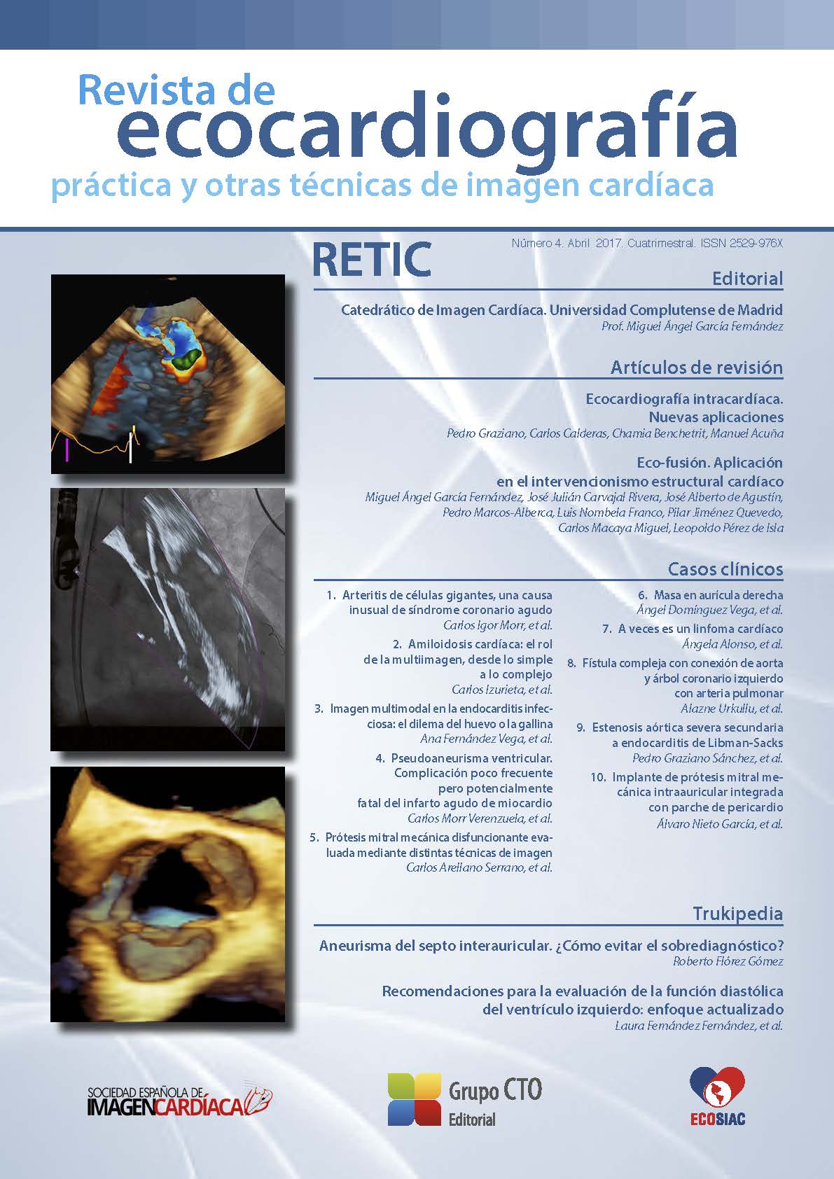
¿Hacia dónde va la ecocardiografía?
DOI:
https://doi.org/10.37615/retic.n4a1Descargas
Métricas
Citas
D’Hooge J, Heimdal A, Jamal F, et al. Regional strain and strain rate measurements by cardiac ultrasound: principles, implementation and limitations. Eur J Echocardiogr 2000; 1: 154-170. DOI: https://doi.org/10.1053/euje.2000.0031
Dandel M, Hetzer R. Echocardiographic strain and strain rate imaging—clinical applications. Int J Cardiol 2009; 132: 11-24. DOI: https://doi.org/10.1016/j.ijcard.2008.06.091
Mor-Avi V, Lang RM, Badano LP, et al. Current and evolving echocardiographic techniques for the quantitative evaluation of cardiac mechanics: ASE/ EAE consensus statement on methodology and indications endorsed by the Japanese Society of Echocardiography. J Am Soc Echocardiogr 2011; 24: 277-313. DOI: https://doi.org/10.1016/j.echo.2011.01.015
Gorcsan J III, Tanaka H. Echocardiographic assessment of myocardial strain. J Am Coll Cardiol 2011; 58: 1401-1413. DOI: https://doi.org/10.1016/j.jacc.2011.06.038
Lang RM, Badano LP, Mor-Avi V, et al. Recommendations for cardiac chamber quantification by echocardiography in adults: an update from the American Society of Echocardiography and the European Association of Cardiovascular Imaging. J Am Soc Echocardiogr 2015; 28: 1-39. DOI: https://doi.org/10.1016/j.echo.2014.10.003
Collier, et al. A Test in Context: Myocardial Strain Measured by Speckle-Tracking Echocardiography. JACC 2017; 69 (8): 1043-1056. DOI: https://doi.org/10.1016/j.jacc.2016.12.012
Thavendiranathan P, Grant AD, Negishi T, et al. Reproducibility of echocardiographic techniques for sequential assessment of left ventricular ejection fraction and volumes: application to patients undergoing cancer chemotherapy. J Am Coll Cardiol 2013; 61 (1): 77-84. DOI: https://doi.org/10.1016/j.jacc.2012.09.035
Vukicevic M, et al. Cardiac 3D Printing and it Future Directions. J Am Coll Cardiol Img 2017; 10 (2): 171-184. DOI: https://doi.org/10.1016/j.jcmg.2016.12.001
Tsang W, Salgo IS, Medvedofsky D, et al. Real-Time Automated Transthoracic Three-Dimensional Echocardiographic Left Heart Chamber Quantification using an Automated Adaptive Analytics Algorithm. JACC Cardiovasc Imaging 2016; 9: 769-782. DOI: https://doi.org/10.1016/j.jcmg.2015.12.020
Arujuna A, Housden R, Ma Y, et al. Novel system for real-time integration of 3D Echocardiography and fluoroscopy for imageguided cardiac interventions: preclinical Validation and clinical feasibility evaluation. IEEE Journal of traslational engineering in health and medicine 2014; 2: 110. DOI: https://doi.org/10.1109/JTEHM.2014.2303799
Balzer J, Zeus T, Hellhammer K, et al. Initial clinical experience using the Echonavigator® – system during structural heart disease interventions. World Journal of Cardiology 2015; 26: 7562-7570. DOI: https://doi.org/10.4330/wjc.v7.i9.562
Feldman T, Hellig F, Mollman H. Structural heart interventions: the state of the art and beyond. Eurointervention 2016; 12: 1-13. DOI: https://doi.org/10.4244/EIJV12SXA1
García-Fernández M, De Agustín A, Pérez de Isla L. Eco-Xray fusion in left atrial appendage closure. Revista Española de Cardiología 2017; 70: 194. DOI: https://doi.org/10.1016/j.rec.2016.05.020
Gaibazzi N, et al. Scar Detection by Pulse-Cancellation Echocardiography: Validation by CMR in Patients With Recent STEMI. JACC Cardiovasc Imaging 2016; 9 :1239-1251. DOI: https://doi.org/10.1016/j.jcmg.2016.01.021
Gupta P, Eisenbrey J, Stanczak M, et al. Effect of Pulse Shaping on Subharmonic Aided Pressure Estimation In Vitro and In Vivo. J Ultrasound Med 2017; 36: 3-11. DOI: https://doi.org/10.7863/ultra.15.11106
Forsberg F, Liu JB, Shi WT, et al. In vivo pressure estimation using subharmonic contrast microbubble signals: proof of concept. IEEE Trans Ultrason Ferroelectr Freq Control 2005; 52: 581-583. DOI: https://doi.org/10.1109/TUFFC.2005.1428040
Dave JK, Halldorsdottir VG, Eisenbrey JR, et al. Noninvasive LV pressure estimation using subharmonic emissions from microbubbles. JACC Cardiovasc Imaging 2012; 5: 87-92.
Dave JK, Halldorsdottir VG, Eisenbrey JR, et al. Subharmonic micro-bubble emissions for noninvasively tracking right ventricular pressures. Am J Physiol Heart Circ Physiol 2012; 303: H126-H132. DOI: https://doi.org/10.1152/ajpheart.00560.2011
Dave JK, Halldorsdottir VG, Eisenbrey JR, et al. Noninvasive LV pressure estimation using subharmonic emissions from microbubbles. JACC Cardiovasc Imaging 2012; 5: 87-92. DOI: https://doi.org/10.1016/j.jcmg.2011.08.017
Descargas
Publicado
Cómo citar
Número
Sección
Licencia
Derechos de autor 2017 Miguel Ángel García Fernández

Esta obra está bajo una licencia internacional Creative Commons Atribución-NoComercial-SinDerivadas 4.0.
RETIC se distribuye bajo la licencia Creative Commons Reconocimiento-NoComercial-SinDerivadas 4.0 Internacional (CC BY-NC-ND 4.0) https://creativecommons.org/licenses/by-nc-nd/4.0 que permite compartir, copiar y redistribuir el material en cualquier medio o formato, bajo los siguientes términos:
- Reconocimiento: debe otorgar el crédito correspondiente, proporcionar un enlace a la licencia e indicar si se realizaron cambios. Puede hacerlo de cualquier manera razonable, pero no de ninguna manera que sugiera que el licenciante lo respalda a usted o su uso.
- No comercial: no puede utilizar el material con fines comerciales.
- No Derivados: si remezcla, transforma o construye sobre el material, no puede distribuir el material modificado.
- Sin restricciones adicionales: no puede aplicar términos legales o medidas tecnológicas que restrinjan legalmente a otros de hacer cualquier cosa que permita la licencia.


