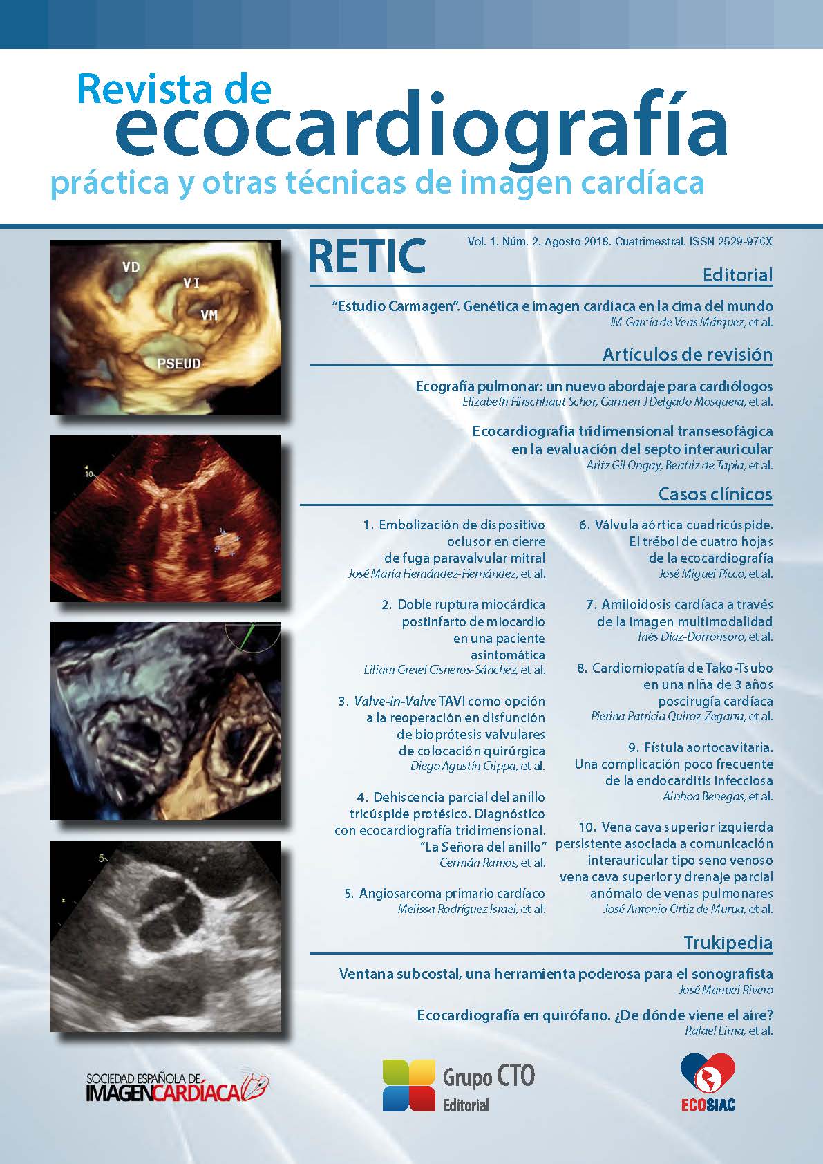
Three-dimensional transesophageal echocardiography in the assessment of the atrial septum
DOI:
https://doi.org/10.37615/retic.v1n2a3Keywords:
3D echocardiography, interatrial septum, patent foramen ovale.Abstract
Transesophageal 3D-echocardiography allows a detailed anatomical observation of the interatrial septum, inclu- ding the remnants of the fetal circulation: the fossa ovalis and the foramen ovale. More than 25% of normal adults present a patent foramen ovale, which under some circumstances may have pathologic relevance. Moreover, most of the structural interventions in the left heart require transseptal crossing of the interatrial septum through the fossa ovalis. Therefore, an adequate knowledge of the anatomical features of the interatrial septum, as well as its normal and pathologic variants is definitely required.
Downloads
Metrics
References
Pushparajah K, Miller OI, Simpson JM. 3D Echocardiography of the Atrial Septum: Anatomical Features and Landmarks for the Echocardiographer. J Am Coll Cardiol Img 2010; 3: 981-984. DOI: https://doi.org/10.1016/j.jcmg.2010.03.015
Faletra FF, Ho SY, Auricchio A. Anatomy of right atrial structures by real-time 3D-transesophageal echocardiography. J Am Coll Cardiol Img 2010; 3 (9): 966-975. DOI: https://doi.org/10.1016/j.jcmg.2010.03.014
Faletra FF, Nucifora G, Ho SY. Imaging the Atrial Septum Using Real-Time Three-Dimensional Transesophageal Echocardiography: Technical Tips, Normal Anatomy, and Its Role in Transseptal Puncture. J Am Soc Echocar- diogr 2011; 24: 593-599. DOI: https://doi.org/10.1016/j.echo.2011.01.022
Silvestry FE, Cohen MS, Armsby LB, et al. Guidelines for the Echocardiographic Assessment of Atrial Septal Defect and Patent Foramen Ovale: From the American Society of Echocardiography and Society for Cardiac Angiography and Interventions. J Am Soc Echocardiogr 2015; 28: 910-958. DOI: https://doi.org/10.1016/j.echo.2015.05.015
Rana BS, Thomas MR, Calvert PA, et al. Echocardiographic Evaluation of Patent Foramen Ovale Prior to Device Closure. J Am Coll Cardiol Img 2010; 3: 749-760. DOI: https://doi.org/10.1016/j.jcmg.2010.01.007
Jensen B, Spicer DE, Sheppard, Anderson RH. Development of the atrial septum in relation to postnatal anatomy and interatrial communications. Heart 2017; 103: 456-462. DOI: https://doi.org/10.1136/heartjnl-2016-310660
Laura DM, Donnino R, Kim E, et al. Lipomatous Atrial Septal Hypertrophy: A Review of Its Anatomy, Pathophysiology, Multimodality Imaging, and Relevance to Percutaneous Interventions. J Am Soc Echocardiogr 2016; 8: 717-723. DOI: https://doi.org/10.1016/j.echo.2016.04.014
Hagen PT, Scholz DG, Edwards WD. Incidence and size of patent foramen ovale during the first 10 decades of life: an autopsy study of 965 normal hearts. Mayo Clin Proc 1984; 59: 17-20. DOI: https://doi.org/10.1016/S0025-6196(12)60336-X
Ho SY, McCarthy KP, Rigby ML. Morphological features pertinent to interventional closure of patent foramen ovale. J Interven Cardiol 2003; 16: 33-38. DOI: https://doi.org/10.1046/j.1540-8183.2003.08000.x
Calvert PA, Rana BS, Kydd AC, Shapiro LM. Patent foramen ovale: anatomy, outcomes, and closure Nat Rev Cardiol 2011; 8: 148-160. DOI: https://doi.org/10.1038/nrcardio.2010.224
Blanche C, Noble S, Roffi M, et al. Platypnea-orthodeoxia syndrome in the elderly treated by percutaneous patent foramen ovale closure: A case series and literature review. Eur J Intern Med 2013; 24: 813-817. DOI: https://doi.org/10.1016/j.ejim.2013.08.698
Bechis MZ, Rubenson DS, Price MJ. Imaging Assessment of the Interatrial Septum for Transcatheter Atrial Septal Defect and Patent Foramen Ovale Closure. Intervent Cardiol Clin 2017; 6: 505-524. DOI: https://doi.org/10.1016/j.iccl.2017.05.004
Downloads
Published
How to Cite
Issue
Section
License
Copyright (c) 2018 Aritz Gil Ongay, Beatriz de Tapia, Juan S Ceña, Iván Olavarri Miguel, José A Vázquez de Prada

This work is licensed under a Creative Commons Attribution-NonCommercial-NoDerivatives 4.0 International License.
RETIC is distributed under the Creative Commons Attribution-NonCommercial-NoDerivatives 4.0 International (CC BY-NC-ND 4.0) license https://creativecommons.org/licenses/by-nc-nd/4.0 which allows sharing, copying and redistribution of the material in any medium or format, under the following terms:
- Attribution: you must give appropriate credit, provide a link to the license, and indicate if changes were made. You may do so in any reasonable manner, but not in any way that suggests that the licensor endorses you or your use.
- Non-commercial: you may not use the material for commercial purposes.
- No Derivatives: if you remix, transform or build upon the material, you may not distribute the modified material.
- No Additional Restrictions: you may not apply legal terms or technological measures that legally restrict others from doing anything permitted by the license.


