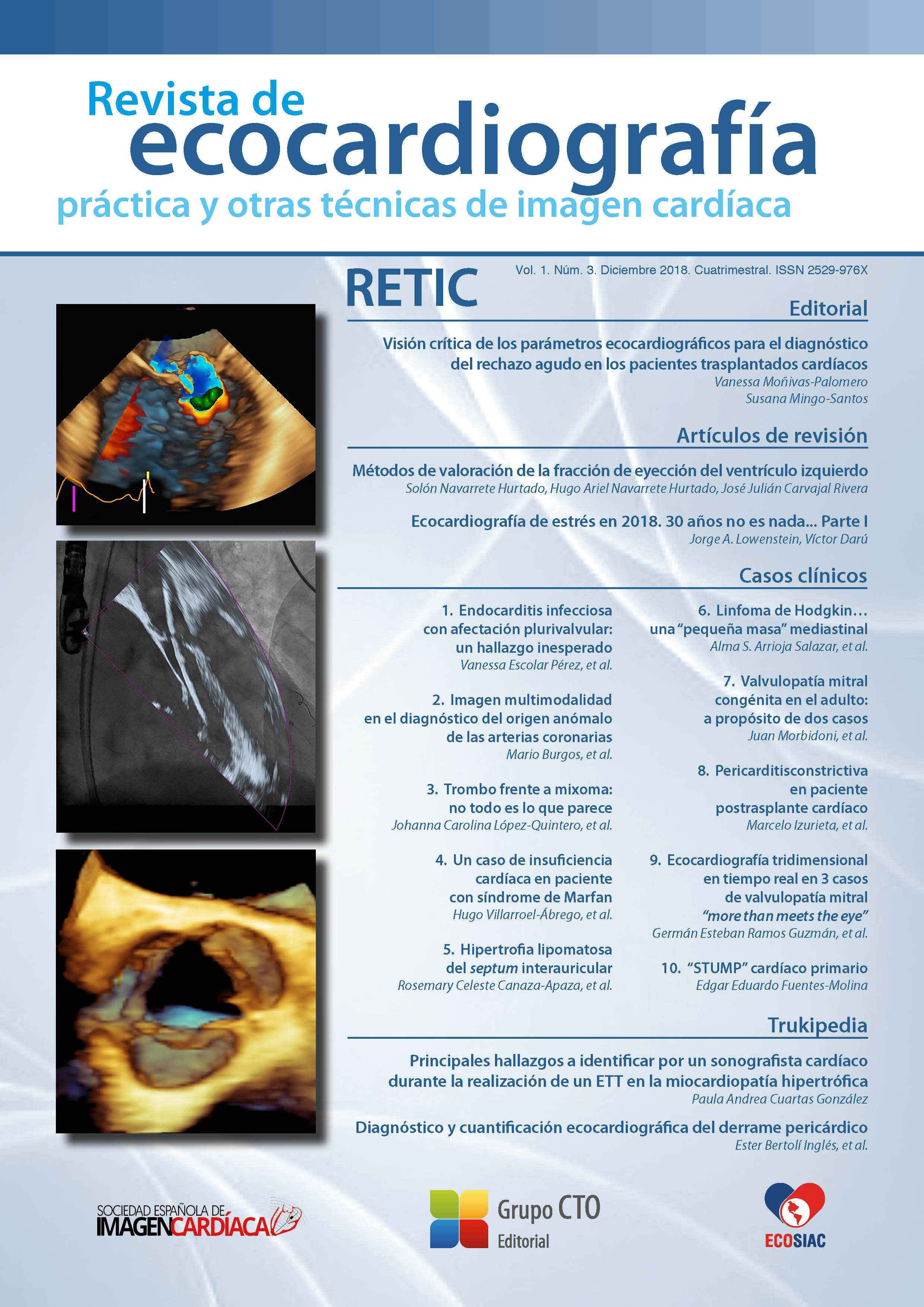
Methods of left ventricular ejection fraction assessment.
DOI:
https://doi.org/10.37615/retic.v1n3a2Keywords:
ventricular function, ejection fraction.Abstract
The ejection fraction is the most widely used index of ventricular function. Its value is pivotal for the clinical decision-making process, including diagnostic, prognostic and therapeutic implications. It is for this reason that the ejection fraction must be an exact, precise measure with minimal uncertainty. In this work we will review the different techniques available for EF calculation, showing their advantages and disadvantages that should be known by the clinician for its proper use.
Downloads
Metrics
References
Ponikowski P, Voor, AA, Anker SD, et al. 2016 ESC Guidelines for the diagnosis and treatment of acute and chronic heart failure: The Task Force for the diagnosis and treatment of acute and chronic heart failure of the European Society of Cardiology (ESC) Developed with the special contribution of the Heart Failure Association (HFA) of the ESC. European Heart Journal, 37 (27), 2.129-2.200.
Zamorano JL, Lancellotti P, Rodriguez Muñoz D, et al. 2016 ESC Position Paper on cancer treatments and cardiovascular toxicity developed under the auspices of the ESC Committee for Practice Guidelines: The Task Force for cancer treatments and cardiovascular toxicity of the European Society of Cardiology (ESC). European Heart Journal, 37 (36), 2.768-2.801.
Keidel WD. U ber eine neue Methode zur Registrierung der Volumen-änderung des Herzens am Menschen. Der Ultraschall in der Medizin, Kongressbericht der Erlangen Ultraschall-Tagung. S. Hirzel Verlag Zürich; 1949. p68-70.
Feigenbaum H, Popp RL, Wolfe SB, et al. Ultrasound measurements of the left ventricle: a correlative study with angiocardiography. Archives of Internal Medicine, 1972, 129 (3), 461-467.
Ahmadpour H, Shah AA, Allen JW, Edmiston WA, Kim SJ, Haywood LJ. Mitral E point septal separation: a reliable index of left ventricular performance in coronary artery disease. American heart journal, 1983, 106 (1), 21-28.
Teichholz LE, Kreulen T, Herman MV, Gorlin R. Problems in echocardiographic volume determinations: echocardiographic-angiographic correlations in the presence or absence of asynergy. The American Journal of Cardiology, 1976, 37 (1), 7-11.
Pai RG, Bodenheimer MM, Pai SM, Koss JH, Adamick RD. Usefulness of systolic excursion of the mitral annulus as an index of left ventricular systolic function. The American Journal of Cardiology, 1991, 67 (2), 222-224.
Fernández MG, Gómez JZ. Procedimientos en ecocardiografía. McGraw-Hill, 2004.
Lang RM, Badano LP, Mor-Avi V, et al. Recommendations for cardiac chamber quantification by echocardiography in adults: an update from the American Society of Echocardiography and the European Association of Cardiovascular Imaging. European Heart Journal-Cardiovascular Imaging, 2015, 16 (3), 233-271.
Von Ramm OT, Smith SW. Real time volumetric ultrasound imaging system. J Digit Imag, 1990. 3: 261-266.
Jenkins C, Bricknell K, Hanekom L, Marwick TH Reproducibility and accuracy of echocardiographic measurements of left ventricular parameters using real-time three-dimensional echocardiography. JACC 2004. 44: 878-886.
Lang RM, Mor-Avi V, Sugeng L. Three-dimensional echocardiography: the benefits of the additional dimension. JACC 2006, 21; 48 (10): 2.053-2.069.
Qin JX, Jones M, Shiota T. Validation of real-time three-dimensional echocardiography for quantifying left ventricular volumes in the presence of a left ventricular aneurysm: in vitro and in vivo studies. JACC 2000. 36: 900-907.
Jenkins C, Bricknell K, Chan J. Comparison of two and three dimensional echocardiography with sequential magnetic resonance imaging for evaluating left ventricular volume and ejection fraction over time in patients with healed myocardial infarction. Am J Cardiol. 2007, 99: 300-306.
Tsang W, Salgo I, Medvedofsky D, et al. Transthoracic 3D echocardiographic left heart chamber quantification using an automated adaptative analytics algorithm. JACC Imaging 2016; 9: 769-782.
Tamborini G, Piazzese C, Lang R, et al. Feasibility and accuracy of automated software for transthoracic three-dimensional left ventricular volume and function analysis: Comparisons with two-dimensional echocardiography, three-dimensional transthoracic manual method, and cardiac magnetic resonance imaging. JASE 2017. Article in press.
Otani K, Nakazono A, Salgo IS. et al. Three-dimensional echocardiographic assessment of left heart chamber size and function with fully automated quantification software in patients with atrial fibrillation. Journal of the American Society of Echocardiography, 2016, 29 (10), 955-965.
Hiremath PS, Akkasaligar PT, Badiger S. Speckle noise reduction in medical ultrasound images. In Advancements and Breakthroughs in Ultrasound Ima- ging. InTech 2013.
Navarrete H. Electromagnetic models for ultrasound image processing. PhD [dissertation]. Universidad Politécnica de Cataluña, 2016. Barcelona. España.
Navarrete S. Un nuevo enfoque para la evaluación de la fracción de eyección. PhD [dissertation]. Universidad Complutense de Madrid, 2017. Madrid. España.
Downloads
Published
How to Cite
Issue
Section
License
Copyright (c) 2018 Solón Navarrete Hurtado, Hugo Ariel Navarrete Hurtado, José Julián Carvajal Rivera

This work is licensed under a Creative Commons Attribution-NonCommercial-NoDerivatives 4.0 International License.
RETIC is distributed under the Creative Commons Attribution-NonCommercial-NoDerivatives 4.0 International (CC BY-NC-ND 4.0) license https://creativecommons.org/licenses/by-nc-nd/4.0 which allows sharing, copying and redistribution of the material in any medium or format, under the following terms:
- Attribution: you must give appropriate credit, provide a link to the license, and indicate if changes were made. You may do so in any reasonable manner, but not in any way that suggests that the licensor endorses you or your use.
- Non-commercial: you may not use the material for commercial purposes.
- No Derivatives: if you remix, transform or build upon the material, you may not distribute the modified material.
- No Additional Restrictions: you may not apply legal terms or technological measures that legally restrict others from doing anything permitted by the license.


