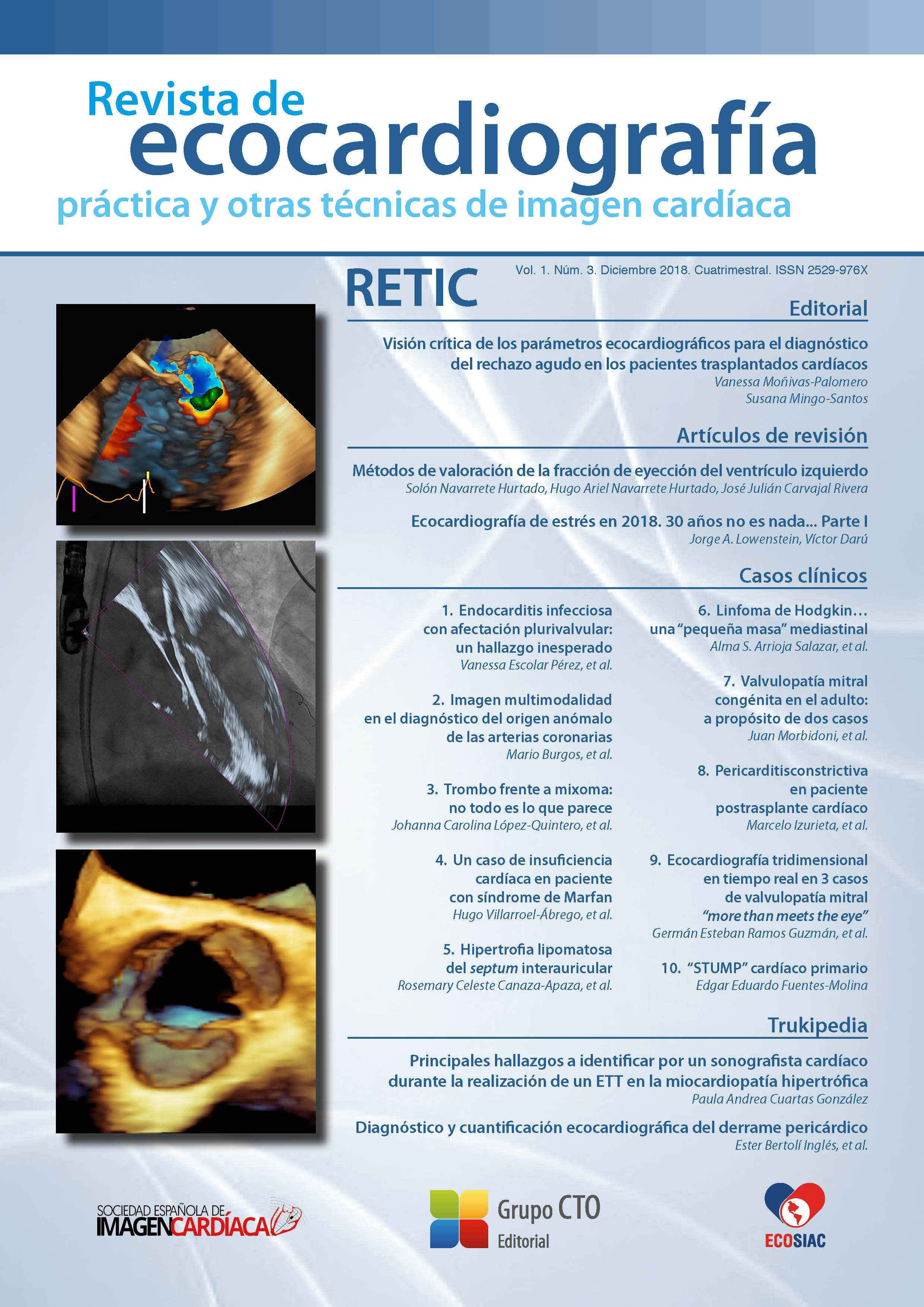
Real-time three-dimensional echocardiography in 3 cases of mitral valve disease "more than meets the eye"
DOI:
https://doi.org/10.37615/retic.v1n3a12Keywords:
three-dimensional transesophageal echocardiography, mitral prosthetic ring thrombosis, mitral valve prolapse, perforated endocarditis.Abstract
Three-dimensional transesophageal echocardiography (TEE-3D) has emerged in recent years as a tool of great assistance to the two-dimensional technique, especially what is related to the study of mitral valve by its location in the near field, which allows an accurate and detailed evaluation of it. We present, through the description 3 cases (prosthetic ring thrombosis, valvular prolapse and perforation by endocarditis) the advantages that the real time 3D-image can offer us in daily practice.
Downloads
Metrics
References
Laplace G, Lafitte S, Labèque J, et al. Clinical significance of early thrombosis after prosthetic mitral valve replacement: a postoperative monocentric study of 680 patients. J Am Coll Cardiol 2004; 43: 1.283-1.290.
Dangas G, Weitz J, Giustino G, et al. Prosthetic Heart Valve Thrombosis. J Am Coll Cardiol 2016; 68: 2.670-2.689.
Ozkan M, Gürsoy OM, Astarcıoğlu MA, et al. Real-time three dimensional transesophageal echocardiography in the assessment of mechanical prosthetic mitral valve ring thrombosis. Am J Cardiol 2013; 1; 112 (7): 977-983.
Gürsoy OM, Karakoyun S, Kalçık M, Özkan M. The incremental value of RT three-dimensional TEE in the evaluation of prosthetic mitral valvering thrombosis complicated with thromboembolism. Echocardiography 2013; 30 (7): E198-201.
Benenstein R, Saric M. Mitral valve prolapse: role of 3D echocardiography in diagnosis. Curr Opin Cardiol 2012, 27: 465-476.
Addetia K, Mor-Avi V, Weinert L, et al. A New Definition for an Old Entity: Improved Definition of Mitral Valve Prolapse Using Three-Dimensional Echocardiography and Color-Coded Parametric Models. J Am Soc Echocardiogr 2014; 27: 8-16.
Faletra F, Demertzis S, Pedrazzini G, et al. Three dimensional transesophageal echocardiography in degenerative mitral regurgitation. J Am Soc Echocardiogr 2015; 28 (4): 437-448.
De Groot-de Laat LE, Ren B, McGhie J, Oei FB, et al. The role of experience in echocardiographic identification of location and extent of mitral valve prolapse with 2D and 3D echocardiography. Int J Cardiovasc Imaging 2016; 32 (8): 1.171-1.177.
Habib G, Hoen B, Tornos P, et al. Guidelines on the prevention, diagnosis, and treatment of infective endocarditis (new version 2009). European Heart Journal 2009; 30 (19): 2.369-2.413.
Salcedo EE, Quaife RA, Seres T, Carroll JD. A framework for systematic characterization of the mitral valve by realtime three-dimensional transesophageal echocardiography. J Am Soc Echocardiogr 2009; 22: 1.087-1.099.
Bhave NM, Addetia K, Spencer KT, et al. Localizing mitral valve perforations with 3D transesophageal echocardiography. JACC Cardiovascular Imaging 2013; 6 (3): 407.
Thompson KA, Shiota T, Tolstrup K, et al. Utility of three-dimensional transesophageal echocardiography in the diagnosis of valvular perforations. American Journal of Cardiology 2011; 107 (1): 100-102.
Downloads
Published
How to Cite
Issue
Section
License
Copyright (c) 2018 Germán Esteban Ramos Guzmán , Manuel Rodríguez Venegas , Mario Zapata Muñoz

This work is licensed under a Creative Commons Attribution-NonCommercial-NoDerivatives 4.0 International License.
RETIC is distributed under the Creative Commons Attribution-NonCommercial-NoDerivatives 4.0 International (CC BY-NC-ND 4.0) license https://creativecommons.org/licenses/by-nc-nd/4.0 which allows sharing, copying and redistribution of the material in any medium or format, under the following terms:
- Attribution: you must give appropriate credit, provide a link to the license, and indicate if changes were made. You may do so in any reasonable manner, but not in any way that suggests that the licensor endorses you or your use.
- Non-commercial: you may not use the material for commercial purposes.
- No Derivatives: if you remix, transform or build upon the material, you may not distribute the modified material.
- No Additional Restrictions: you may not apply legal terms or technological measures that legally restrict others from doing anything permitted by the license.


