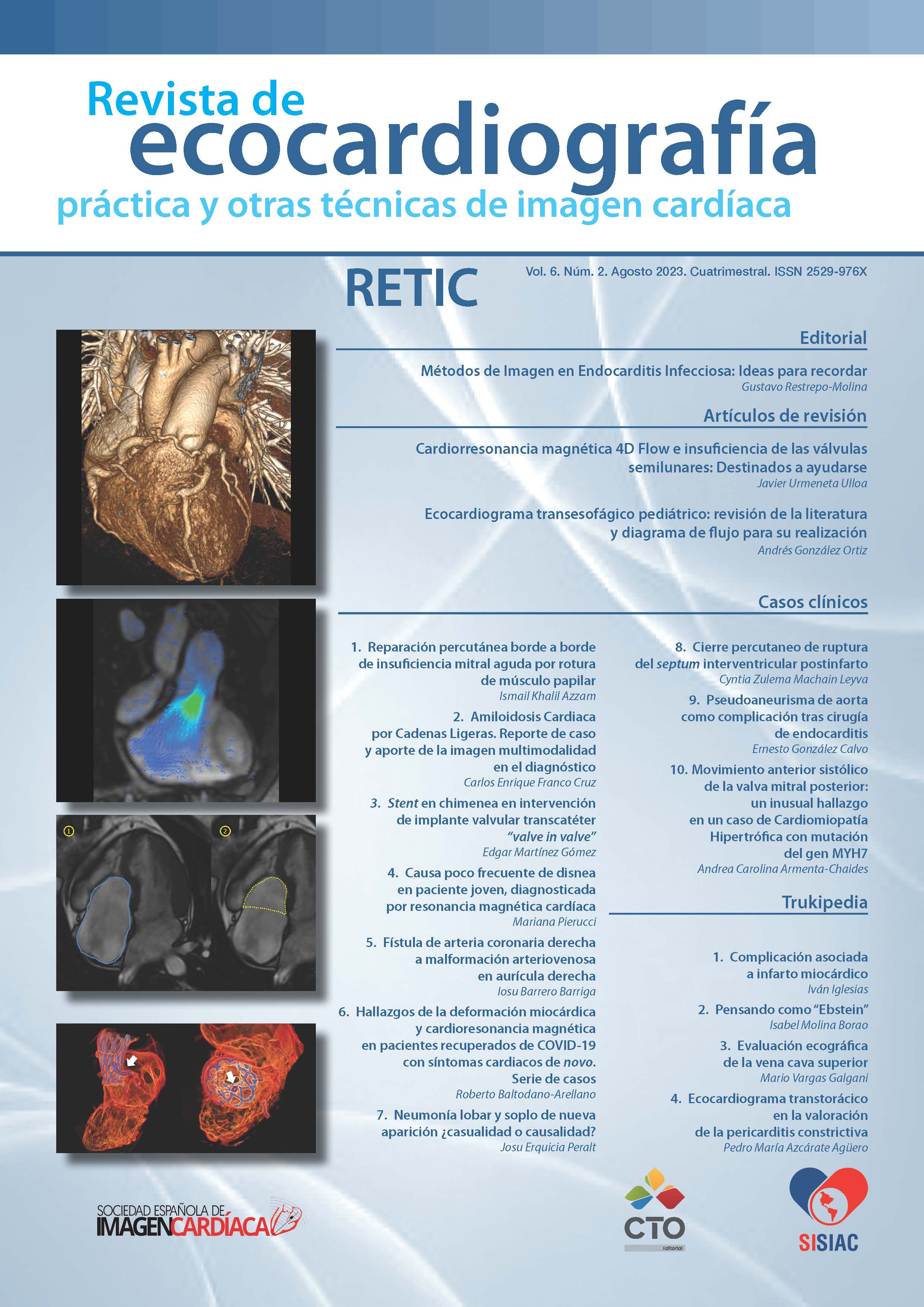
Echographic evaluation of Superior cava vein.
DOI:
https://doi.org/10.37615/retic.v6n2a16Keywords:
echography, superior cava vein, Doppler echoAbstract
Recommendations on ultrasound evaluation of the superior vena cava and its flow, as well as patterns of physiological and pathological variations are presented.
Downloads
Metrics
References
Fadel B, Kazzy B, Mohty D. Ultrasound image of the superior vena cava. J Am Soc Echocardiogr. 2023; 36: 447-463. doi: https://doi.org/10.1016/j.echo.2023.01.017
Sivaciyan V, Ranganathan N. Transcutaneous Doppler jugular venous flow velocity recording. 1978; 57:930-939. doi: https://doi.org/10.1161/01.cir.57.5.930
Murayama M, Kaga S, Okada K. et al. Clinical utility of superior vena cava flow velocity waveform measured from the subcostal window for estimating right atrial pressure. J Am Soc Echocardiogr. 2022; 35:727-737. doi:https://doi.org/10.1016/j.echo.2022.02.002
Downloads
Published
How to Cite
Issue
Section
License
Copyright (c) 2023 Mario Vargas Galgani

This work is licensed under a Creative Commons Attribution-NonCommercial-NoDerivatives 4.0 International License.
RETIC is distributed under the Creative Commons Attribution-NonCommercial-NoDerivatives 4.0 International (CC BY-NC-ND 4.0) license https://creativecommons.org/licenses/by-nc-nd/4.0 which allows sharing, copying and redistribution of the material in any medium or format, under the following terms:
- Attribution: you must give appropriate credit, provide a link to the license, and indicate if changes were made. You may do so in any reasonable manner, but not in any way that suggests that the licensor endorses you or your use.
- Non-commercial: you may not use the material for commercial purposes.
- No Derivatives: if you remix, transform or build upon the material, you may not distribute the modified material.
- No Additional Restrictions: you may not apply legal terms or technological measures that legally restrict others from doing anything permitted by the license.


