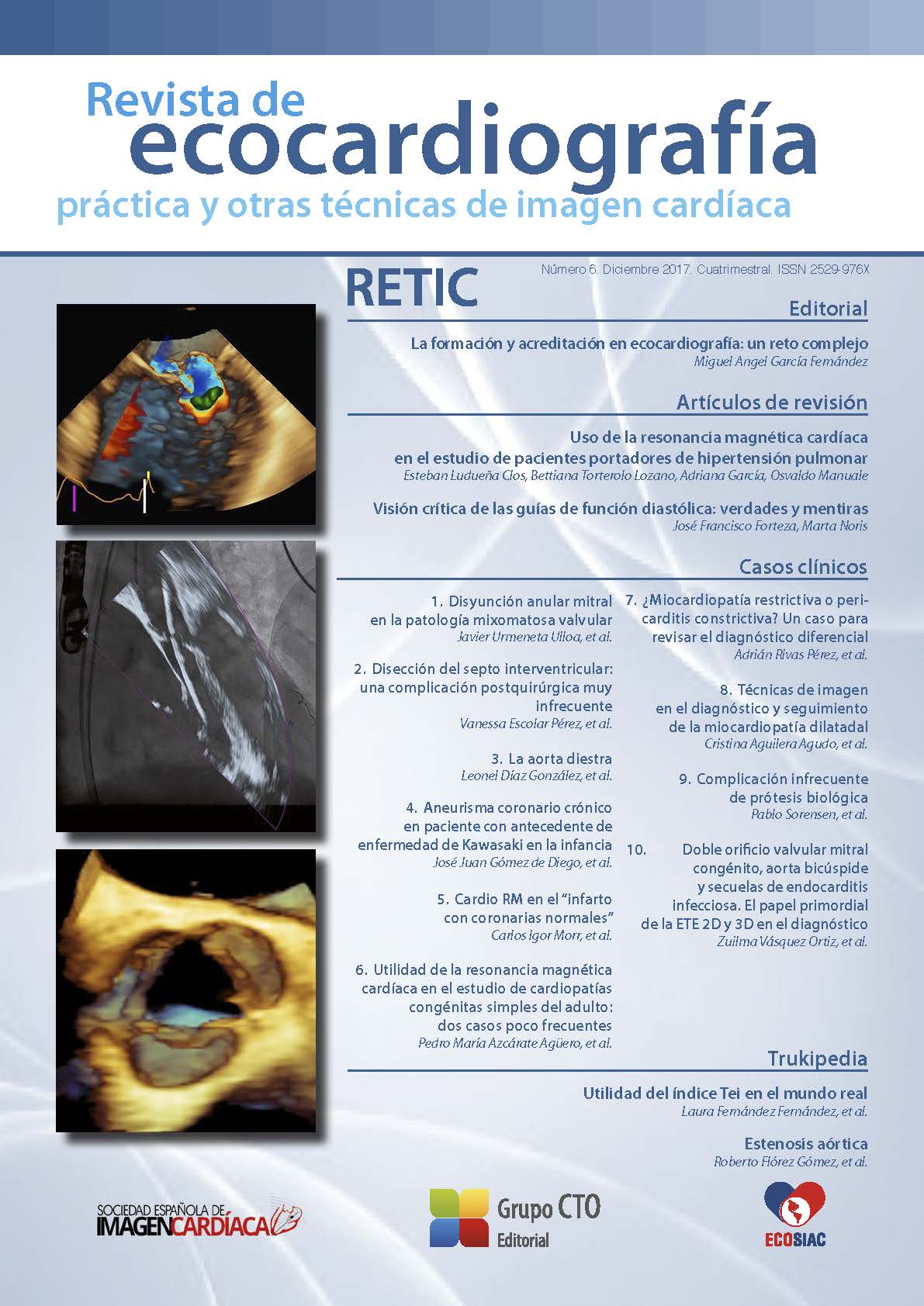
Use of cardiac magnetic resonance imaging in the study of patients with pulmonary hypertension
DOI:
https://doi.org/10.37615/retic.n6a2Keywords:
cardiac magnetic resonance, pulmonary hypertension, late enhancement, myocardial strain.Abstract
Cardiac Magnetic Resonance allows the anatomical and functional assessment of left and right cardiac chambers, and aorto-pulmonary vascular tree in patients with pulmonary hypertension. That information has a high prognostic value previous and during treatment. The protocol has a large number of sequences to anatomical and functional assessment, for myocardial strain by tagging and feature tracking, phase contrast for aortic and pulmonary flows and after the administration of gadolinium, for delayed enhancement and tridimensional reconstruction of the vascular tree.
Downloads
Metrics
References
Pamboucas C, Nihoyannopoulos P. Papel de la resonancia magnética cardiovascular en el diagnóstico y evaluación de la hipertensión pulmonar. Rev Esp Cardiol. 2006; 59 (8): 755-760. DOI: https://doi.org/10.1157/13091877
Grothues F, Moon JC, Bellenger NG, Smith G, Klein H, Pennel D. Interstudy reproducibility of right ventricular volumes, function, and mass with cardiovascularmagnetic resonance. Am Heart J. 2004; 147: 218-222. DOI: https://doi.org/10.1016/j.ahj.2003.10.005
Lorenz CH, Walker ES, Morgan VI, Klein SS, Graham Jr TP. Normal humanright and left ventricular mass, systolic function, and gender differences by cine magnetic resonance imaging. J Cardiovasc Magn Reson. 1999; 1: 7-21. DOI: https://doi.org/10.3109/10976649909080829
Mackey ES, Sandler MP, Campbel RM; Graham TP Jr, Atkinson JB, Prie R, Moreau GA: Right ventricular myocardial mass quantification with magnetic resonance imaging. Am J Cardiol 1990; 65: 529-532. DOI: https://doi.org/10.1016/0002-9149(90)90828-O
Boxt LM, Katz J: Magnetic resonance imaging for quantitation of right ventricular volumen in patients with pulmonary hypertension. J Thorac Imaging 1993; 8: 92-97. DOI: https://doi.org/10.1097/00005382-199321000-00002
Raymond RJ, Hinderliter AL, Willis PW, Ralph D, Caldwell EJ, Williams W, Ettinger NA, Hill NS, Summer Wr, de Boisblanc B, et al. Echocardiographic predictors of adverse otucomes in primary pulmonary hypertension. J Am Coll Cardiol 2002; 39: 1.214:1.219. DOI: https://doi.org/10.1016/S0735-1097(02)01744-8
Heath D, Edwards JE: Configuration of elastic tissue of pulmonary trunk in idiopathic pulmonary hypertension. Circulation 1960; 21: 59-62. DOI: https://doi.org/10.1161/01.CIR.21.1.59
Wilkins M, Paul G, Strange J, Tunariu N, Gin-Sing W, Banya W, et al. Sildenafil versus endothelin receptor antagonist for pulmonary hypertension (SERAPH) study. Am J Respir Crit Care Med. 2005; 171: 1.292-1.297. DOI: https://doi.org/10.1164/rccm.200410-1411OC
Heiberg E, Sjögren J, Ugander M, Carlsson M, Engblom H, and Arheden H. Design and Validation of Segment – a Freely Available Software for Cardiovascular Image Analysis, BMC Medical Imaging, 2010, 10: 1. DOI: https://doi.org/10.1186/1471-2342-10-1
Bradlow WM, Hughes M, Keen NG, Bucciarelli-Ducci C, Assomull R, Gibbs JS, Mohiaddin RH. Measuring the heart in pulmonary arterial hypertension (PAH): implications for trial study size. J Magn Reson Imaging 2010; 31: 117-124. DOI: https://doi.org/10.1002/jmri.22011
Saba TS, Foster J, Cockburn M, Cowan M, Peacock AJ. Ventricular mass index using magnetic resonance imaging accurately estimates pulmonary artery pressure. Eur Respir J. 2002; 20: 1.519-1.524. DOI: https://doi.org/10.1183/09031936.02.00014602
Roeleveld RJ, Marcus JT, Faes TJ, Gan TJ, Boonstra A. Postmus PE, et al. Interventricular septal configuration at MR imaging and pulmonary arterial pressure in pulmonary hypertension. Radiology. 2005; 234: 710-717. DOI: https://doi.org/10.1148/radiol.2343040151
Haber I, Metaxas D, Geval T, Axel L. Three-dimensional systolic kinematics of the right ventricle. Am J Physiol Heart Circ Physiol. 2005; 289: H1.826-1.833. DOI: https://doi.org/10.1152/ajpheart.00442.2005
Fayad ZA, Ferrari VA, Kraitchman DL, Young AA, Palevsky HI, Bloomgarden DC, Axel L. Right ventricular regional function usin MR tagging: normals versus chronic pulmonary hypertension. Magn Reson Med 1998; 39: 116-123. DOI: https://doi.org/10.1002/mrm.1910390118
Marcus JT, Gan CT, Zwanenburg JJ, Boonstra A, Allaart CP, Gotte MJ, Vank-Noordegraaf A: Interventricular mechanical asynchrony in pulmonary arterial hypertension: left-to-right delay in peak shortening is realted to rightventricular overload and left ventricular underfilling. J Am Coll Cardiol 2008; 51: 750-757. DOI: https://doi.org/10.1016/j.jacc.2007.10.041
Mousseaux E, Tasu JP, Jolivet O, Simonneau G, Bittoun J, Gaux JC. Pulmonary arterial resistance: noninvasive measurement with indexes of pulmonary flow estimated at velocity-encoded MR imaging-preliminary experience. Radiology. 1999; 212: 896-902. DOI: https://doi.org/10.1148/radiology.212.3.r99au21896
Sanz J, Kuschnir P, Rius T, Salguero R, Sulica R, Einstein AJ, Dellegrottaglie S, Fuster V, Rajagopalan S, Poon M: Pulmonary arterial hypertension: noninvasive detection with phase-constrast MR imaging. Radiology 2007; 243: 70-79. DOI: https://doi.org/10.1148/radiol.2431060477
Kind T, Maurtiz GI, Marcus JT, van de Veerdonk M, Westerhof N, Vank-Noordegraaf : Right ventricular ejection fraction is better reflected by transverse rather tan longitudinal wal motion in pulmonary hypertension. J Cardiovasc Magn Reson 2010; 12: 35. DOI: https://doi.org/10.1186/1532-429X-12-35
Pamboucas C, Schmitz S, Nihoyannopoulos P. Magnetic resonance imaging in the detection of myocardial viability: te role of delayed contrast hypernancement. Ellenic J Cardiol. 2005; 46: 108-116.
Blyth K, Groenning B, Martin T, Foster JE, Mark PB, Dargie HJ, et al. Contrast enhanced-cardiovascular magnetic resonance imaging in patients with pulmonary hypertension. Eur Heart J. 2005; 26: 1.993-1.999. DOI: https://doi.org/10.1093/eurheartj/ehi328
Kruger S, Haage P, Hoffmann R, Breuer C, Bucker A, Hanrath P, et al. Diagnosis of pulmonary arterial hypertension and pulmonary embolism with magnetic resonance angiopraphy. Chest. 2001; 120: 156-161. DOI: https://doi.org/10.1378/chest.120.5.1556
Levin D, Hatabu H. MR Evaluation of pulmonary blood flow. J Thorac Imaging. 2004; 19: 241-249. DOI: https://doi.org/10.1097/01.rti.0000142836.14611.f2
Torbicki A. Cardiac magnetic resonance in pulmonary arterial hypertension: a step in the right direction. Eur Heart J. 2007; 28: 1.187-1.189. DOI: https://doi.org/10.1093/eurheartj/ehm074
Downloads
Published
How to Cite
Issue
Section
License
Copyright (c) 2017 Esteban Ludueña Clos, Bettiana Torterolo Lozano, Adriana García, Osvaldo Manuale

This work is licensed under a Creative Commons Attribution-NonCommercial-NoDerivatives 4.0 International License.
RETIC is distributed under the Creative Commons Attribution-NonCommercial-NoDerivatives 4.0 International (CC BY-NC-ND 4.0) license https://creativecommons.org/licenses/by-nc-nd/4.0 which allows sharing, copying and redistribution of the material in any medium or format, under the following terms:
- Attribution: you must give appropriate credit, provide a link to the license, and indicate if changes were made. You may do so in any reasonable manner, but not in any way that suggests that the licensor endorses you or your use.
- Non-commercial: you may not use the material for commercial purposes.
- No Derivatives: if you remix, transform or build upon the material, you may not distribute the modified material.
- No Additional Restrictions: you may not apply legal terms or technological measures that legally restrict others from doing anything permitted by the license.


