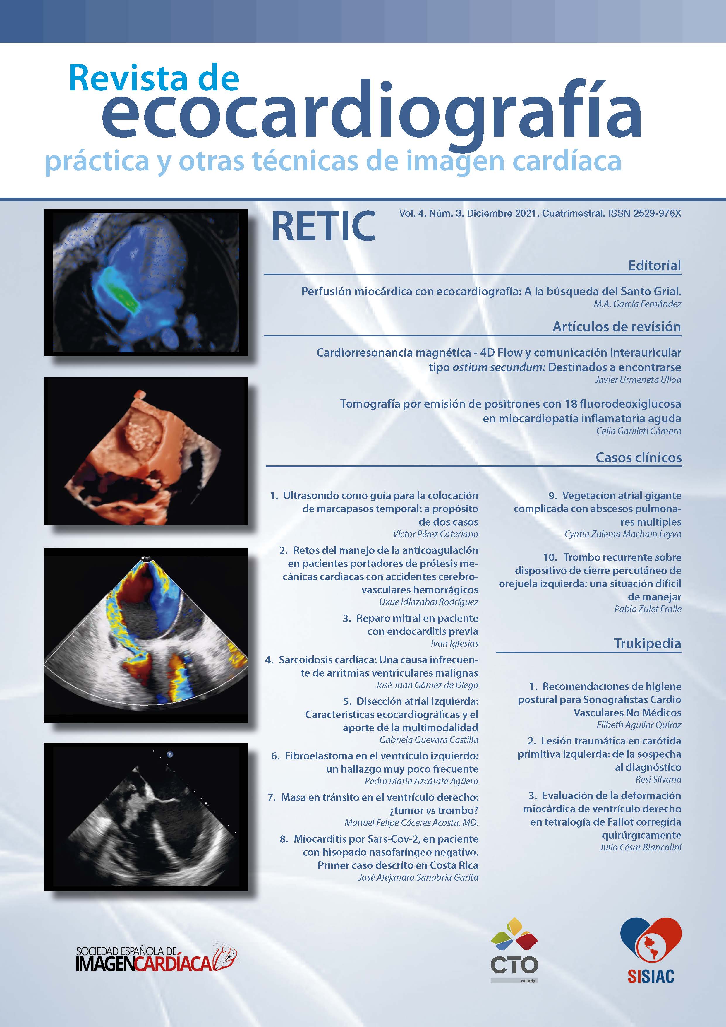
Perfusión miocárdica con ecocardiografía: A la búsqueda del Santo Grial
DOI:
https://doi.org/10.37615/retic.v4n3a1Descargas
Métricas
Citas
Gramiak R, Shah PM. Echocardiography of the aortic root. Invest Radiol. 1968 Sep-Oct;3(5):356-66.
Feinstein SB, Ten Cate FJ, Zwehl W, Ong K, Maurer G, Tei C, et al. Two-dimensional contrast echocardiography. I. In vitro development and quantitative analysis of echo contrast agents. J Am Coll Cardiol. 1984 Jan;3(1):14-20.
Serra V, García Fernández M, Zamorano J. Microbubbles: basic principles. Contrast echocardiography in Clinical Practice Springer Verlag; 2004. p. 19-44. 4. Cheng SC, Dy TC, Feinstein SB. Contrast echocardiography: review and future directions. Am J Cardiol. 1998 Jun 18;81(12A):41G-8G.
Garcia-Fernandez MA, Macchioli RO, Moreno PM, Yanguela MM, Thomas JB, Sendon JL, et al. Use of contrast echocardiography in the diagnosis of subacute myocardial rupture after myocardial infarction. J Am Soc Echocardiogr. 2001 Sep;14(9):945-7.
Waggoner AD, Williams GA, Gaffron D, Schwarze M. Potential utility of left heart contrast agents in diagnosis of myocardial rupture by 2-dimensional echocardiography. J Am Soc Echocardiogr. 1999 Apr;12(4):272-4.
Nakatani S, Imanishi T, Terasawa A, Beppu S, Nagata S, Miyatake K. Clinical application of transpulmonary contrast-enhanced Doppler technique in the assessment of severity of aortic stenosis. J Am Coll Cardiol. 1992 Oct;20(4):973-8.
Ha JW, Lee BK, Kim HJ, Pyun WB, Byun KH, Rim SJ, et al. Assessment of left atrial appendage filling pattern by using intravenous administration of microbubbles: comparison between mitral stenosis and mitral regurgitation. J Am Soc Echocardiogr. 2001 Nov;14(11):1100-6.
Fernandez Portales J, Garcia Fernandez MA, Moreno M, Gonzalez Alujas MT, Placer JL, Allue C, et al. [Usefulness of the new imaging techniques, second harmonic and contrast in endocardial border visualization. Reliability analysis in segmental contraction assessment]. Rev Esp Cardiol. 2000 Nov;53(11):1459-66.
Reilly JP, Tunick PA, Timmermans RJ, Stein B, Rosenzweig BP, Kronzon I. Contrast echocardiography clarifies uninterpretable wall motion in intensive care unit patients. J Am Coll Cardiol. 2000 Feb;35(2):485-90.
Yong Y, Wu D, Fernandes V, Kopelen HA, Shimoni S, Nagueh SF, et al. Diagnostic accuracy and cost-effectiveness of contrast echocardiography on evaluation of cardiac function in technically very difficult patients in the intensive care unit. Am J Cardiol. 2002 Mar 15;89(6):711-8.
Kusnetzky LL, Khalid A, Khumri TM, Moe TG, Jones PG, Main ML. Acute mortality in hospitalized patients undergoing echocardiography with and without an ultrasound contrast agent: results in 18,671 consecutive studies. J Am Coll Cardiol. 2008 Apr 29;51(17):1704-6.
Thomson HL, Basmadjian AJ, Rainbird AJ, Razavi M, Avierinos JF, Pellikka PA, et al. Contrast echocardiography improves the accuracy and reproducibility of left ventricular remodeling measurements: a prospective, randomly assigned, blinded study. J Am Coll Cardiol. 2001 Sep;38(3):867-75.
Lepper W, Hoffmann R, Kamp O, Franke A, Cock CC, Kühl HP, Sieswerda GT, Dahl JV, Janssens U, Voci P Visser CA, Hanrath P. Assessment of myocardial reperfusion by intravenous myocardial contrast echocardiography and coronary flow reserve after primary percutaneous transluminal coronary angiography in patients with acute myocardial infarction. Circulation 2000; 101: 2368-2374.
Jayaweera AR, Edwards N, Glasheen WP, Villanueva FS, Abbott RD, Kaul S. In vivo myocardial kinetics of air-filled albumin microbubbles during myocardial contrast echocardiography. Comparison with radiolabeled red blood cells. Circulation Research 1994; 74: 1157-1165.
Jayaweera AR, Matthew TL, Sklenar J, Spotnitz WD, Watson DD, Kaul S. Method for the quantitation of myocardial perfusion during myocardial contrast two-dimensional echocardiography. J Am Soc Echocardiogr 1990;3(2):91-98.
Cheirif J, Zoghbi WA, Raizner AE, Minor ST, Winters WL Jr, Klein MS, De Bauche TL, Lewis JM, Roberts R, Quiñones MA. Assessment of myocardial perfusion in humans by contrast echocardiography. I. Evaluation of regional coronary reserve by peak contrast intensity. J Am Coll Cardiol. 1988;11(4): 735-743.
Amarjit J , Michael Hickman, O Kamp, R M. Lang, J D. Thomas, M A. Vannan, Je Vanoverschelde, Poll A. Van Der Wouw, Roxy Senior*Myocardial contrast echocardiography for the detection of coronary artery stenosis - A prospective multicenter study in comparison with single-photon emission computed tomographyJournal of the American College of Cardiology 47(1):141-5
Senior R. Moreo A. Gaibazzi N. Agati L. Tiemann K. Shivalkar B. et al. Comparison of sulfur hexafluoride microbubble (SonoVue)-enhanced myocardial echocardiography to gated single photon emission computerized tomography for the detection of significant coronary artery disease: a large European multicentre study. J Am Coll Cardiol. 2013; 62: 1353-1361
Rinkevich D, Kaul S, Wang XQ, Tong KL, Belcik T, Kalvaitis S, et al. Regional left ventricular perfusion and function in patients presenting to the emergency department with chest pain and no ST-segment elevation. Eur Heart J. 2005 Aug;26(16):1606-11.
Rezkalla SH, Kloner RA. No-reflow phenomenon. Circulation. 2002 Feb 5;105(5):656-62. 22. Perez David E, Garcia Fernandez MA. Myocardial contrast echocardiography in acute myocardial infarction: the importance of assessing coronary microcirculation. Rev Esp Cardiol. 2004 Jan;57(1):4-6.
Perez-David E, Garcia-Fernandez MA, Quiles J, Mahia P, Lopez-Sendon JL, Lopez de Sa E, et al. Usefulness of quantitative myocardial contrast echocardiography for prediction of ventricular function recovery after myocardial infarction treated with primary angioplasty. Heart. 2006 May;92(5):693-4.
Tardif JC, Dore A, Chan KL, Fagan S, Honos G, Marcotte F, et al. Economic impact of contrast stress echocardiography on the diagnosis and initial treatment of patients with suspected coronary artery disease. J Am Soc Echocardiogr. 2002 Nov;15(11):1335-45.
Moir S, Haluska BA, Jenkins C, Fathi R, Marwick TH. Incremental benefit of my cardial contrast to combined dipyridamole-exercise stress echocardiography for the assessment of coronary artery disease. Circulation. 2004 Aug 31;110(9):1108-13.
Elhendy A, O’Leary EL, Xie F, McGrain AC, Anderson JR, Porter TR. Comparative accuracy of real-time myocardial contrast perfusion imaging and wall motion analysis during dobutamine stress echocardiography for the diagnosis of coronary artery disease. J Am Coll Cardiol. 2004 Dec 7;44(11):2185-91.
Masugata H, Peters B, Lafitte S, Strachan M, Ohmori K, DeMaria AN. Quantitative assessment of myocardial perfusion during graded coronary stenosis by real-time myocardial contrast echo refilling curves. J Am Coll Cardiol 2001; 37: 262-269.
Lafitte S, Higashiyama A, Masugata H, Peters B, Strachan M, Kwan OL, DeMaria AN. Contrast echocardiography can assess risk area and infarct size during coronary occlusion and reperfusion: experimental validation. J Am Coll Cardiol 2002; 39: 1546-1554.
Porter TR, Xie F, Kricsfeld A, Kilzer K. Noninvasive identification of acute myocardial ischemia and reperfusion with contrast ultrasound using intravenous perfluoropropane-exposed sonicated dextrose albumin. J Am Coll Cardiol 1995;26(1):33-40.
Meza M, Greener Y, Hunt R, Perry B, Revall S, Barbee W, et al. Myocardial contrast echocardiography: reliable, safe, and efficacious myocardial perfusion assessment after intravenous injections of a new echocardiographic contrast agent. Am Heart J 1996;132(4):871-881.
Coggins MP, Sklenar J, Le DE, Wei K, Lindner JR, Kaul S. Noninvasive prediction of ultimate infarct size at the time of acute coronary occlusion based on the extent and magnitude of collateral-derived myocardial blood flow. Circulation 2001;104(20):2471-2477.
Kaul S, Glasheen W, Ruddy TD, Pandian NG, Weyman AE, Okada RD. The importance of defining left ventricular area at risk in vivo during acute myocardial infarction: an experimental evaluation with myocardial contrast two-dimensional echocardiography. Circulation. 1987;75(6):1249-1260.
GarciaFernandez MA, Zamorano J.Contrat Echocardiography in clinical Practice.Spring Verlag Berlin 2004.
Descargas
Publicado
Cómo citar
Número
Sección
Licencia
Derechos de autor 2021 Miguel Ángel García Fernández

Esta obra está bajo una licencia internacional Creative Commons Atribución-NoComercial-SinDerivadas 4.0.
RETIC se distribuye bajo la licencia Creative Commons Reconocimiento-NoComercial-SinDerivadas 4.0 Internacional (CC BY-NC-ND 4.0) https://creativecommons.org/licenses/by-nc-nd/4.0 que permite compartir, copiar y redistribuir el material en cualquier medio o formato, bajo los siguientes términos:
- Reconocimiento: debe otorgar el crédito correspondiente, proporcionar un enlace a la licencia e indicar si se realizaron cambios. Puede hacerlo de cualquier manera razonable, pero no de ninguna manera que sugiera que el licenciante lo respalda a usted o su uso.
- No comercial: no puede utilizar el material con fines comerciales.
- No Derivados: si remezcla, transforma o construye sobre el material, no puede distribuir el material modificado.
- Sin restricciones adicionales: no puede aplicar términos legales o medidas tecnológicas que restrinjan legalmente a otros de hacer cualquier cosa que permita la licencia.


