
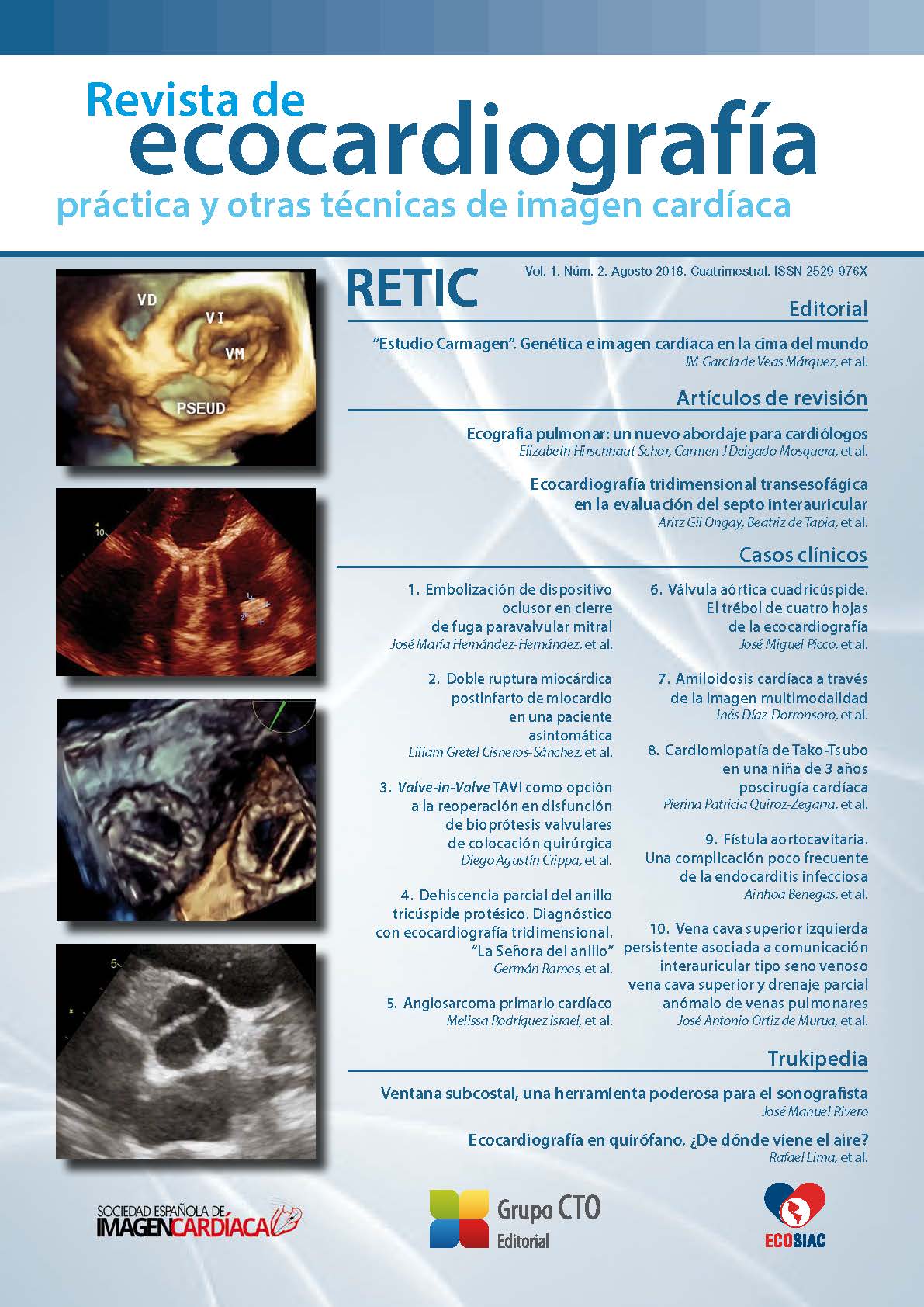
Dehiscencia parcial del anillo tricúspide protésico. Diagnóstico con ecocardiografía tridimensional. “La Señora del anillo”
DOI:
https://doi.org/10.37615/retic.v1n2a7Palabras clave:
dehiscencia parcial de anillo protésico tricúspide, insuficiencia tricúspide, ecocardiografía tridimensional.Resumen
Una mujer de 58 años con antecedentes de reemplazo valvular mitral mecánico y anuloplastia con anillo protésico tricúspide desarrolló un cuadro de deterioro de la capacidad funcional y edema de las extremidades inferiores. La ecocardiografía transtorácica mostró dilatación del ventrículo derecho con función sistólica preservada e insuficiencia tricúspide (IT) excéntrica grave con un mecanismo poco claro. La ecocardiografía transesofágica confirmó estos hallazgos y reveló una dehiscencia parcial del anillo protésico. La reconstrucción tridimensional en face de la válvula tricúspide permitió la caracterización del defecto y el desarrollo de una estrategia adecuada por el equipo quirúrgico.
Existe evidencia creciente de la presencia de insuficiencia tricúspide residual significativa postoperatoria. La dehiscencia del anillo protésico es un mecanismo infrecuente que eventualmente podría requerir una nueva operación. Por ello, su diagnóstico es relevante y las técnicas novedosas como la ecocardiografía tridimensional pueden ayudar significativamente en la toma de decisiones.
Descargas
Métricas
Citas
Rodés-Cabau J, Taramasso M, O’Gara PT. Diagnosis and treatment of tricuspid valve disease: current and future perspectives. Lancet 2016; 388: 2431- 2442. DOI: https://doi.org/10.1016/S0140-6736(16)00740-6
Wang N, Phan S, Tian DH, Yan TD, Phan K. Flexible band versus rigid ring annuloplasty for tricuspid regurgitation: a systematic review and meta-analysis. Ann Cardiothorac Surg 2017; 6 (3): 194-203. DOI: https://doi.org/10.21037/acs.2017.05.05
Parolari A, Barili F, Pilozzi A, Pacini D. Ring or suture annuloplasty for tricuspid regurgitation? A meta-analysis review. Ann Thorac Surg 2014; 98: 2255-2263. DOI: https://doi.org/10.1016/j.athoracsur.2014.06.100
Pfannmüller B, Doenst T, Eberhardt K, et al. Increased risk of dehiscence after tricuspid valve repair with rigid annuloplasty rings. J Thorac Cardiovasc Surg 2012; 143 (5): 1050-1055. DOI: https://doi.org/10.1016/j.jtcvs.2011.06.019
Tei C, Pilgrim JP, Shah PM, et al. The tricuspid valve annulus: study of size and motion in normal subjects and in patients with tricuspid regurgitation. Circulation 1982; 66: 665-671. DOI: https://doi.org/10.1161/01.CIR.66.3.665
Ton-Nu TT, Levine RA, Handschumacher MD, et al. Geometric determinants of functional tricuspid regurgitation: insights from 3-dimensional echocardiography. Circulation 2006; 114: 143-149. DOI: https://doi.org/10.1161/CIRCULATIONAHA.106.611889
Paul DM, Naran A, Pierce EL, et al. Suture dehiscence in the Tricuspid Annulus: An Ex Vivo Analysis of Tissue Strength and Composition. Ann Thorac Surg 2017; 104 (3): 820-826. DOI: https://doi.org/10.1016/j.athoracsur.2017.02.040
Pfannmuller B, Davierwala P, Misfeld M, et al. Postoperative outcome of isolated tricuspid valve operation using arrested-heart or beating heart technique. Ann Thorac Surg 2012; 94: 1218-1222. DOI: https://doi.org/10.1016/j.athoracsur.2012.05.020
Miglioranza MH, Mihaila S, Muraru D, et al. Dynamic changes in tricuspid annular diameter measurement in relation to the echocardiographic view and timing during the cardiac cycle. J Am Soc Echocardiogr 2015; 28: 226- 235. DOI: https://doi.org/10.1016/j.echo.2014.09.017
Shiota T. Role of modern 3D echocardiography in valvular heart disease. Korean J Intern Med 2014; 29 (6): 685-702. DOI: https://doi.org/10.3904/kjim.2014.29.6.685
Naqvi TZ, Rafie R, Ghalichi M. Real-time 3D TEE for the diagnosis of rightsided endocarditis in patients with prosthetic devices. JACC Cardiovasc Imaging 2010; 3 (3): 325-327. DOI: https://doi.org/10.1016/j.jcmg.2009.11.011
Sugeng L, Shernan SK, Weinert L, et al. Real-time three-dimensional transesophageal echocardiography in valve disease: comparisonwith surgical findings and evaluation of prosthetic valves. J Am Soc Echocardiog 2008; 21 (12): 1347-1354. DOI: https://doi.org/10.1016/j.echo.2008.09.006
Urmeneta Ulloa J, Molina Borao I, Aured Guallar C, et al. Three-Dimensional Echocardiography in the Evaluation of the Dehiscence of Mitral Valve Annuloplasty Ring. Circulation 2015; 132 (25): e388-390. DOI: https://doi.org/10.1161/CIRCULATIONAHA.115.016064
Begüm Uygur, Mehmet Ertürk, Hale Ünal Aksu, Aydın Yıldırım. Partial detachment of tricuspid valve annuloplasty ring detected by three-dimensional transesophageal Echocardiography. Anatol J Cardiol 2016; 7118: E 9. DOI: https://doi.org/10.14744/AnatolJCardiol.2016.7118
Descargas
Publicado
Cómo citar
Número
Sección
Licencia
Derechos de autor 2018 Germán Ramos, Mario Zapata, Manuel Rodríguez, Sebastián Herrera

Esta obra está bajo una licencia internacional Creative Commons Atribución-NoComercial-SinDerivadas 4.0.
RETIC se distribuye bajo la licencia Creative Commons Reconocimiento-NoComercial-SinDerivadas 4.0 Internacional (CC BY-NC-ND 4.0) https://creativecommons.org/licenses/by-nc-nd/4.0 que permite compartir, copiar y redistribuir el material en cualquier medio o formato, bajo los siguientes términos:
- Reconocimiento: debe otorgar el crédito correspondiente, proporcionar un enlace a la licencia e indicar si se realizaron cambios. Puede hacerlo de cualquier manera razonable, pero no de ninguna manera que sugiera que el licenciante lo respalda a usted o su uso.
- No comercial: no puede utilizar el material con fines comerciales.
- No Derivados: si remezcla, transforma o construye sobre el material, no puede distribuir el material modificado.
- Sin restricciones adicionales: no puede aplicar términos legales o medidas tecnológicas que restrinjan legalmente a otros de hacer cualquier cosa que permita la licencia.


