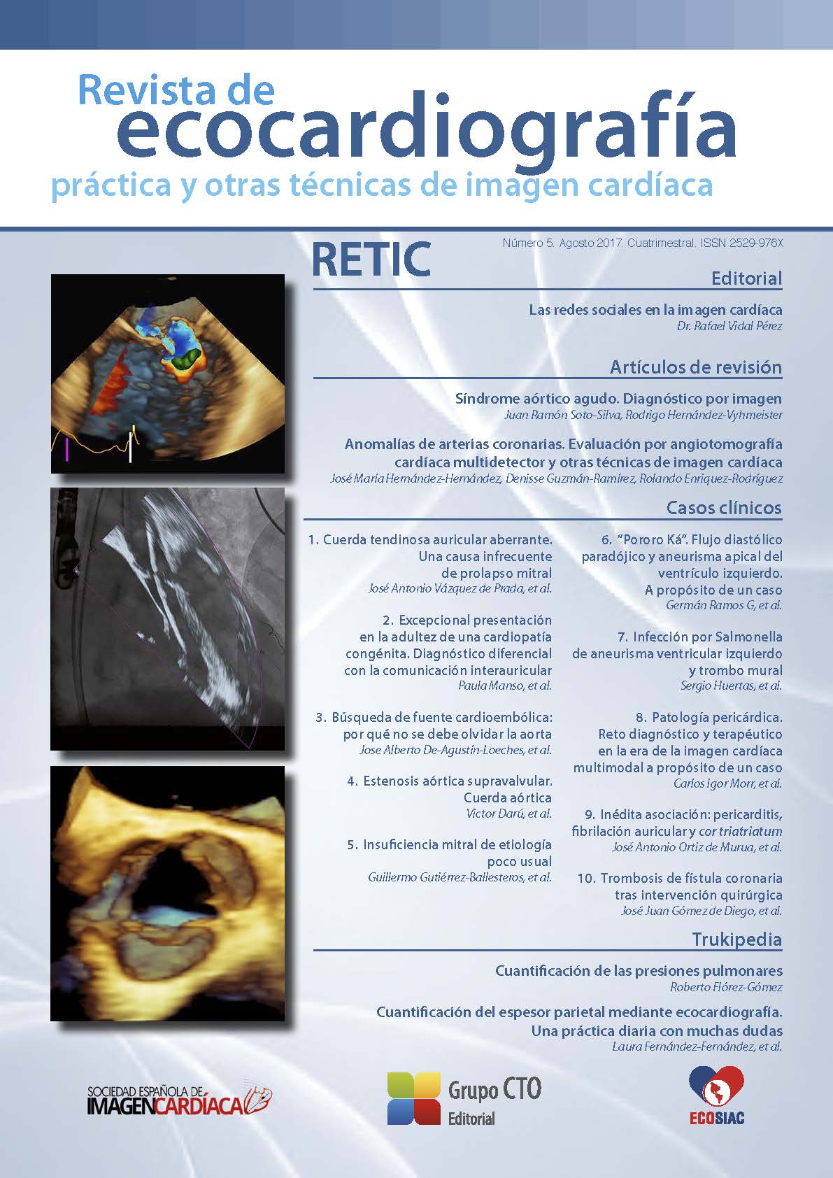
Coronary artery anomalies. Evaluation by multidetector cardiac angiotomography and other cardiac imaging techniques
DOI:
https://doi.org/10.37615/retic.n5a3Keywords:
coronary artery anomalies, multidetector computed tomography-coronary angiography, sudden cardiac death, cardiac imaging techniques.Abstract
Coronary artery anomalies occur in 1.7% of the general population and cause 33% of sudden deaths in young people during strenuous exertion. The use of cardiac imaging techniques has allowed us to expand our knowledge in the diagnosis of these anomalies. There are three types according to Greenberg classification, origin, course and termination anomalies. The most important are those with hemodynamic compromise which are coronary atresia, ALCAPA/ARCAPA, L-ACAOS, R-ACAOS, coronary artery fistulae and coronary artery ectasias. The evaluation by multidetector computed tomography-coronary angiography allows us to characterize them from the ostium to its termination and to demonstrate the clinical consequences in the patient, other imaging techniques such as stress cardiac magnetic resonance imaging, stress echocardiography, myocardial perfusion single photon emission computed tomography and coronary angiography with evaluation by coronary reserve flow or free-wave instantaneous radio allow selection of appropriate treatment.
Downloads
Metrics
References
Angelini P. Novel imaging of coronary artery anomalies to assess their prevalence, the causes of clinical symptoms, and the risk of sudden cardiac death. Circ Cardiovasc Imaging 2014; 7: 747-754.
Angelini P, Villason S, Cha AV, Diez JG. Normal and anomalous coronary arteries in humans. En: Angelini P (ed.). Coronary Artery Anomalies: A Comprehensive Approach. Lippincott Williams and Wilkins. Philadelphia, 1999; 27-79.
Muriago M, Sheppard M, Ho S, Anderson R. The location of the coronary arterial orifices in the normal heart. Clin Anat 1997; 10: 1-6. DOI: https://doi.org/10.1002/(SICI)1098-2353(1997)10:5<297::AID-CA1>3.0.CO;2-O
Jacobs JE. Computed tomographic evaluation of the normal cardiac anatomy. Radiol Clin North Am 2010; 48: 701-710. DOI: https://doi.org/10.1016/j.rcl.2010.05.001
Erol C, Şeker M. The prevalence of coronary artery variations on coronary computed tomography angiography. Acta Radiol 2012; 53: 278-284. DOI: https://doi.org/10.1258/ar.2011.110394
Schlesinger MI. Relation of anatomic pattern to pathologic condition of the coronary arteries. Arch Pathol 1940; 30: 403-415.
Kini S, Bis KG, Weaver L. Normal and variant coronary arterial and venous anatomy on high-resolution CT angiography. AJR Am J Roentgenol 2007; 188: 1665-1674. DOI: https://doi.org/10.2214/AJR.06.1295
Cademartiri F, La Grutta L, Malag R, et al. Prevalence of anatomical variants and coronary anomalies in 543 consecutive patients studied with 64-slice CT coronary angiography. Eur Radiol 2008; 18: 781-791. DOI: https://doi.org/10.1007/s00330-007-0821-9
Erol C, Koplay M, Paksoy Y. Evaluation of anatomy, variation and anomalies of the coronary arteries with coronary computed tomography angiography. Anadolu Kardyol Derg 2013; 13: 154-164. DOI: https://doi.org/10.5152/akd.2013.041
Angelini P. Coronary artery anomalies: an entity in search of identity. Circulation 2007; 115 (10): 1296-1305. DOI: https://doi.org/10.1161/CIRCULATIONAHA.106.618082
Chen HI, Poduri A, Numi H, et al. VEGF-C and aortic cardiomyocytes guide coronary artery stem development. J Clin Invest 2014; 124: 4899-4914. DOI: https://doi.org/10.1172/JCI77483
Pérez-Pomares JM, de la Pompa JL, Franco D, et al. Congenital coronary artery anomalies: a bridge from embriology to anatomy and pathophisiology-a position statement of the development, anatomy, and pathology ESC Working Group. Cardiovasc Res 2016; 109: 204-216. DOI: https://doi.org/10.1093/cvr/cvv251
Risau W, Flamme I. Vasculogenesis. Annu Rev Cell Biol Dev Biol 1995; 11:73-91. DOI: https://doi.org/10.1146/annurev.cb.11.110195.000445
Angelini P, Velasco JA, Flamm S. Coronary anomalies: incidence, pathophysiology, and clinical relevance. Circulation 2002; 105 (20): 2449-2454. DOI: https://doi.org/10.1161/01.CIR.0000016175.49835.57
Greenberg MA, Fish BG, Spindola-Franco H. Congenital anomalies of the coronary arteries. Classification and significance. Radiol Clin North Am 1989; 27 (6): 1127-1146.
Angelini P. Novel Imaging of Coronary Artery Anomalies to Assess Their Prevalence, the Causes of Clinical Symptoms, and the Risk of Sudden Cardiac Death. Circ Cardiovasc Imaging 2014; 7 (4): 747-754. DOI: https://doi.org/10.1161/CIRCIMAGING.113.000278
Zeina AR, Blinder J, Sharif D, et al. Congenital coronary artery anomalies in adults: non-invasive assessment with multidetector CT. The British Journal of Radiology 2009; 82: 254-261. DOI: https://doi.org/10.1259/bjr/80369775
Sundaram B, Kreml R, Patel S. Imaging of coronary artery anomalies. Radiol Clin North Am 2010; 48: 711-727. DOI: https://doi.org/10.1016/j.rcl.2010.04.006
Koşar P, Ergün E, .Ztürk C, Koşar U. Anatomic variations and anomalies of the coronary arteries: 64-slice CT angiographic appearance. Diagn Interv Radiol 2009; 15: 275-283. DOI: https://doi.org/10.4261/1305-3825.DIR.2550-09.1
Erol C, Şeker M. Coronary artery anomalies: the prevalence of origination, course, and termination anomalies of coronary arteries detected by 64-detector computed tomography coronary angiography. J Comput Assist Tomogr 2011; 35: 618-624. DOI: https://doi.org/10.1097/RCT.0b013e31822aef59
Lipton MJ, Barry WH, Obrez I, et al. Isolated single coronary artery: diagnosis, angiographic classification, and clinical significance. Radiology 1979; 130: 39-47. DOI: https://doi.org/10.1148/130.1.39
Wesselhoeft H, Fawcett JS, Johnson AL. Anomalous origin of the left coronaryartery from the pulmonary trunk: its clinical spectrum, pathology and pathophysiology, based on review of 140 cases with seven further cases. Circulation 1968; 38: 403-425. DOI: https://doi.org/10.1161/01.CIR.38.2.403
Waite S, Ng T, Afari A, Gohari A, Lowery R. CT diagnosis of isolated anomalous origin of the RCA arising from the main pulmonary artery. J Thorac Imaging 2008; 23: 145-147. DOI: https://doi.org/10.1097/RTI.0b013e3181653c5a
Yau JM, Singh R, Halpern EJ, Fischman D. Anomalous origin of the left coronary artery from the pulmonary artery in adults: a comprehensive review of 151 adult cases and a new diagnosis in a 53-year-old woman. Clin Cardiol 2011; 34 (4): 204-210. DOI: https://doi.org/10.1002/clc.20848
Basso C, Maron BJ, Corrado D, Thiene G. Clinical profile of congenital coronary artery anomalies with origin from the wrong aortic sinus leading to sudden death in young competitive athletes. J Am Coll Cardiol 2000; 35:1493-1501. DOI: https://doi.org/10.1016/S0735-1097(00)00566-0
Cheezum MK, Ghoshhajra B, Bittencourt MS, et al. Anomalous origin of the coronary artery arising from the opposite sinus: prevalence and outcomes in patients undergoing coronary CTA. EHJ Cardiovasc Imag 2017; 18: 224- 235. DOI: https://doi.org/10.1093/ehjci/jev323
Gebauer R, Cerny S, Vojtovic P, Tax P. Congenital atresia of the left coronary artery--myocardial revascularization in two children. Interact Cardiovasc Thorac Surg 2008; 7: 1174-1175. DOI: https://doi.org/10.1510/icvts.2008.184317
Mohlenkamp S, Hort W, Ge J, Erbel R. Update on myocardial bridging. Circulation 2002; 106: 2616-2622. DOI: https://doi.org/10.1161/01.CIR.0000038420.14867.7A
Tarantini G, Migliore F, Cademartiri F, et al. Left anterior descending artery myocardial bridging. JACC 2016; 68 (25): 2887-2899. DOI: https://doi.org/10.1016/j.jacc.2016.09.973
Antoniadis AP, Chatzizisis YS, Giannoglou GD. Pathogenetic mechanisms of coronary ectasia. Int J Cardiol 2008; 130 (3): 335-343. DOI: https://doi.org/10.1016/j.ijcard.2008.05.071
Warnes C, Williams RG, Bashore TM, et al. ACC/AHA 2008 guidelines for the management of adults with congenital heart disease: a report of the American College of Cardiology/American Heart Association Task Force on Practice Guidelines (Writing Committee to Develop Guidelines on the Management of Adults with Congenital Heart Disease. J Am Coll Cardiol 2008; 52: e143-e263.
Saboo SS, Juan YH, Khandelwal A, et al. MDCT of congenital coronary artery fistulas. ARJ 2014; 203: W244-W252. DOI: https://doi.org/10.2214/AJR.13.12026
Moberg A. Anastomoses between extracardiac vessels and coronary arteries. I. Via bronchial arteries: post-mortem angiographic study in adults and newborn infants. Acta Radiol Diagn 1967; 6 (2): 177-192. DOI: https://doi.org/10.1177/028418516700600209
Downloads
Published
How to Cite
Issue
Section
License
Copyright (c) 2017 José María Hernández-Hernández, Denisse Guzmán-Ramírez, Rolando Enriquez-Rodríguez

This work is licensed under a Creative Commons Attribution-NonCommercial-NoDerivatives 4.0 International License.
RETIC is distributed under the Creative Commons Attribution-NonCommercial-NoDerivatives 4.0 International (CC BY-NC-ND 4.0) license https://creativecommons.org/licenses/by-nc-nd/4.0 which allows sharing, copying and redistribution of the material in any medium or format, under the following terms:
- Attribution: you must give appropriate credit, provide a link to the license, and indicate if changes were made. You may do so in any reasonable manner, but not in any way that suggests that the licensor endorses you or your use.
- Non-commercial: you may not use the material for commercial purposes.
- No Derivatives: if you remix, transform or build upon the material, you may not distribute the modified material.
- No Additional Restrictions: you may not apply legal terms or technological measures that legally restrict others from doing anything permitted by the license.


