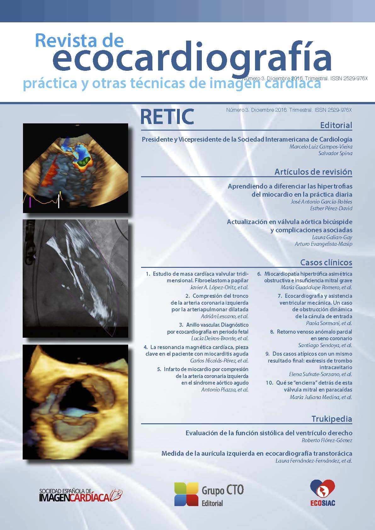
Learning to differentiate myocardial hypertrophies in daily practice
DOI:
https://doi.org/10.37615/retic.n3a2Keywords:
ventricular hypertrophy, echocardiography, cardiac magnetic resonance imaging.Abstract
Left ventricular hypertrophy is frequently seen in ecochardiographic laboratories and everyone knows that there are many etiologies involved in this condition. This is why we need keys that help us to diagnose the cause. It is true that there are not pathognomonic facts for every disease, but we can suspect the etiology using all the data collected in each patient, basically coming from echocardiography and cardiac magnetic resonance (CMR). In this article we sumarize the available information for pathologic conditions (hypertrophic cardiomyopathy, hypertensive cardiopathy, infiltrative diseases like amioloidosis and storage diseases like Fabry disease) and the heart of the athlete. The last one have been choosed due to, taht in some instances is an important differential diagnosis with a state of disease.
Downloads
Metrics
References
Elliott P, McKenna WJ. Hypertrophic cardiomyopathy. Lancet 2004; 363: 1.881-1.891. DOI: https://doi.org/10.1016/S0140-6736(04)16358-7
Weideman F, Niemann M, Ertl G, Störk S. The Different Faces of Echocardiographic left Ventricular Hypertrophy: Clues to the Etiology. J Am Soc Echocardiogr 2010; 23: 793-801. DOI: https://doi.org/10.1016/j.echo.2010.05.020
Janardhanan R, Kramer CM. Imaging in hypertensive heart disease. Expert Rev Cardiovasc Ther 2011; 9: 199-209. DOI: https://doi.org/10.1586/erc.10.190
Palmon LC, Reichek N, Yeon SB, et al. Intramural myocardial shortening in hypertensive left ventricular hypertrophy with normal pump function. Circulation 1994; 89: 122-131. DOI: https://doi.org/10.1161/01.CIR.89.1.122
Rudolph A, Abdel-Aty H, Bohl S, et al. Noninvasive detection of fibrosis applying contrast enhanced cardiac magnetic resonance in different forms of left ventricular hypertrophy relation to remodeling. J Am Coll Cardiol 2009; 53: 284-291. DOI: https://doi.org/10.1016/j.jacc.2008.08.064
Kim JH, Baggish AL. Differentiating Exercise-Induced Cardiac Adaptations From Cardiac Pathology: The “Grey Zone” of Clinical Uncertainty. Canadian J Cardiol 2016; 32: 429-437. DOI: https://doi.org/10.1016/j.cjca.2015.11.025
La Gerche A, Baggish AL, Knuuti J, et al. Cardiac Imaging and Stress Testing Asymptomatic Athletes to Identify Those at Risk of Sudden Cardiac Death. J Am Coll Cardiol Img 2013; 6: 993-1.007. DOI: https://doi.org/10.1016/j.jcmg.2013.06.003
Elliott PM, Anastasakis A, Borger MA, et al. 2014 ESC Guidelines on diagnosis and management of hypertrophic cardiomyopathy: The Task Force for the Diagnosis and Management of Hypertrophic Cardiomyopathy of the European Society of Cardiology (ESC). Eur Heart J 2014; 35: 2.733- 2.779. DOI: https://doi.org/10.1093/eurheartj/ehu284
Sherrid MV, Balaram S, Kim B, et al. The Mitral Valve in Obstructive Hypertrophic Cardiomyopathy: A Test in Context. J Am Coll Cardiol 2016; 67: 1.846- 1.858. DOI: https://doi.org/10.1016/j.jacc.2016.01.071
Kato TS, Noda A, Izawa H, et al. Discrimination of nonobstructive hypertrophic cardiomyopathy from hypertensive left ventricular hypertrophy on the basis of strain rate imaging by tissue Doppler ultrasonography. Circulation 2004; 110: 3.808-3.814. DOI: https://doi.org/10.1161/01.CIR.0000150334.69355.00
Rickers C, Wilke NM, Jerosch-Herold M, et al. Utility of cardiac magnetic resonance imaging in the diagnosis of hypertrophic cardiomyopathy. Circulation 2005; 112: 855-861. DOI: https://doi.org/10.1161/CIRCULATIONAHA.104.507723
Ho CY, Abbasi SA, Neilan TG, et al. T1 measurements identify extracellular volume expansion in hypertrophic cardiomyopathy sarcomere mutation carriers with and without left ventricular hypertrophy. Circ Cardiovasc Imaging 2013; 6: 415-422. DOI: https://doi.org/10.1161/CIRCIMAGING.112.000333
González-López E, Gallego-Delgado M, Guzzo-Merello G, et al. Wild-type transthyretin amyloidosis as a cause of heart failure with preserved ejection fraction. Eur Heart J 2015; 36: 2.585-2.594. DOI: https://doi.org/10.1093/eurheartj/ehv338
Sun JP, Stewart WJ, Yang XS, et al. Differentiation of hypertrophic cardiomyopathy and cardiac amyloidosis from other causes of ventricular wall thickening by two-dimensional strain imaging echocardiography. Am J Cardiol 2009; 103: 411-415. DOI: https://doi.org/10.1016/j.amjcard.2008.09.102
Espinosa MA, Pérez David E, Carrillo R, et al. Los criterios ecocardiográficos son insuficientes para establecer un diagnóstico precoz de amiloidosis: estudio comparativo con RM cardiaca. Rev Esp Cardiol 2015; 68 (1): 317.
Maceira AM, Joshi J, Prasad SK, et al. Cardiovascular magnetic resonance in cardiac amyloidosis. Circulation 2005; 111: 186-193. DOI: https://doi.org/10.1161/01.CIR.0000152819.97857.9D
Fontana M, Banypersad SM, Treibel TA, et al. Differential Myocyte Responses in Patients with Cardiac Transthyretin Amyloidosis and Light-Chain Amyloidosis: A Cardiac MR Imaging Study. Radiology 2015; 277: 388-397. DOI: https://doi.org/10.1148/radiol.2015141744
Perugini E, Guidalotti PL, Salvi F, et al. Noninvasive etiologic diagnosis of cardiac amyloidosis using 99mTc-3,3- diphosphono-1,2-propanodicarboxylic acid scintigraphy. J Am Coll Cardiol 2005; 46: 1.076-1.084. DOI: https://doi.org/10.1016/j.jacc.2005.05.073
Seydelmann N, Wanner C, Störk S, et al. Fabry Disease and the Heart. Best Practice & Research Clinical Endocrinology & Metabolism 2015; 29: 195-204. DOI: https://doi.org/10.1016/j.beem.2014.10.003
Cardiovascular magnetic resonance demonstration of the spectrum of morphological phenotypes and patterns of myocardial scarring in Anderson-Fabry disease. J Cardiovasc Magn Reson 2016; 18: 14-23. DOI: https://doi.org/10.1186/s12968-016-0233-6
Pica S, Sado DM, Maestrini V, et al. Reproducibility of native myocardial T1 mapping in the assessment of Fabry disease and its role in early detection of cardiac involvement by cardiovascular magnetic resonance. J Cardiovasc Magn Reson 2014; 16: 99-107. DOI: https://doi.org/10.1186/s12968-014-0099-4
Downloads
Published
How to Cite
Issue
Section
License
Copyright (c) 2016 José Antonio García-Robles , Esther Pérez-David

This work is licensed under a Creative Commons Attribution-NonCommercial-NoDerivatives 4.0 International License.
RETIC is distributed under the Creative Commons Attribution-NonCommercial-NoDerivatives 4.0 International (CC BY-NC-ND 4.0) license https://creativecommons.org/licenses/by-nc-nd/4.0 which allows sharing, copying and redistribution of the material in any medium or format, under the following terms:
- Attribution: you must give appropriate credit, provide a link to the license, and indicate if changes were made. You may do so in any reasonable manner, but not in any way that suggests that the licensor endorses you or your use.
- Non-commercial: you may not use the material for commercial purposes.
- No Derivatives: if you remix, transform or build upon the material, you may not distribute the modified material.
- No Additional Restrictions: you may not apply legal terms or technological measures that legally restrict others from doing anything permitted by the license.


