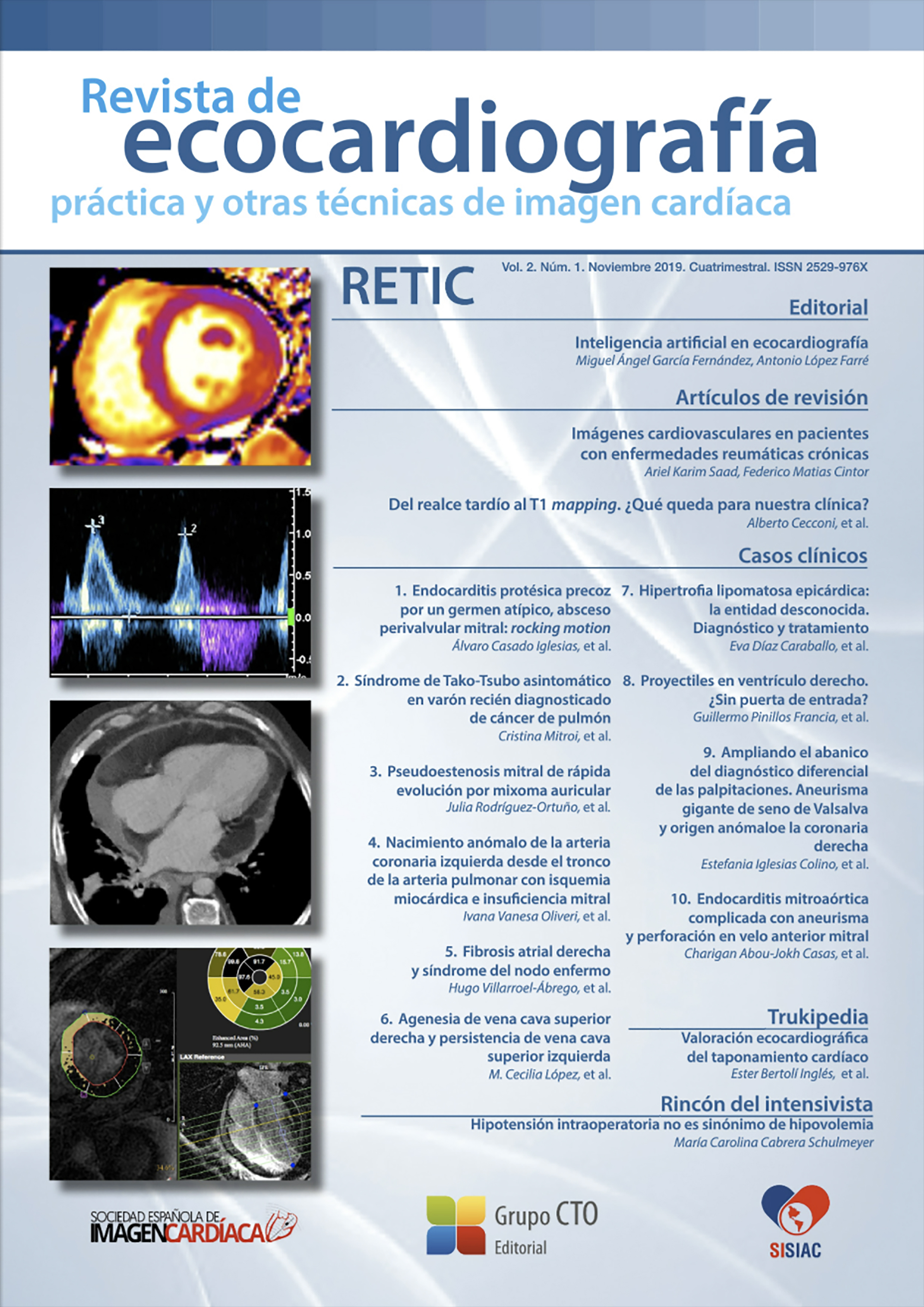
From late enhancement to T1 mapping. What is left for our clinic?
DOI:
https://doi.org/10.37615/retic.v2n1a3Keywords:
myocardial fibrosis, late gadolinium enhancement, T1 mapping, extracellular volume.Abstract
Myocardial fibrosis is a pathological process common to most of heart diseases. However, the type of myocardial involvement can differ significantly depending on its etiology. Tissue characterization of myocardial fibrosis patterns can be explored in a complementary way by the sequences of late gadolinium enhancement and T1 mapping. In this review, we will discuss current evidence for the use of these imaging techniques and describe their most prominent clinical applications.
Downloads
Metrics
References
Perea Palazon RJ, Ortiz Perez JT, Prat Gonzalez S, de Caralt Robira TM, Cibeira Lopez MT, Sole Arques M. Parametric techniques for characterizing myocardial tissue by magnetic resonance imaging (part 1): T1 mapping. Radiologia. 2016;58(3):164-77. doi: https://doi.org/10.1016/j.rx.2015.12.007
Ambale-Venkatesh B, Lima JA. Cardiac MRI: a central prognostic tool in myocardial fibrosis. Nature reviews Cardiology. 2015;12(1):18-29. doi: https://doi.org/10.1038/nrcardio.2014.159
Haaf P, Garg P, Messroghli DR, Broadbent DA, Greenwood JP, Plein S. Cardiac T1 Mapping and Extracellular Volume (ECV) in clinical practice: a comprehensive review. Journal of cardiovascular magnetic resonance : official journal of the Society for Cardiovascular Magnetic Resonance. 2016;18(1):89. doi: https://doi.org/10.1186/s12968-016-0308-4
Satoh H, Sano M, Suwa K, Saitoh T, Nobuhara M, Saotome M, et al. Distribution of late gadolinium enhancement in various types of cardiomyopathies: Significance in differential diagnosis, clinical features and prognosis. World journal of cardiology. 2014;6(7):585-601. doi: https://doi.org/10.4330/wjc.v6.i7.585
Windecker S, Kolh P, Alfonso F, Collet JP, Cremer J, Falk V, et al. 2014 ESC/EACTS guidelines on myocardial revascularization. EuroIntervention : journal of EuroPCR in collaboration with the Working Group on Interventional Cardiology of the European Society of Cardiology. 2015;10(9):1024-94. doi: https://doi.org/10.4244/EIJY14M09_01
Romero J, Xue X, Gonzalez W, Garcia MJ. CMR imaging assessing viability in patients with chronic ventricular dysfunction due to coronary artery disease: a meta-analysis of prospective trials. JACC Cardiovascular imaging. 2012;5(5):494-508. doi: https://doi.org/10.1016/j.jcmg.2012.02.009
Bekkers SC, Yazdani SK, Virmani R, Waltenberger J. Microvascular obstruction: underlying pathophysiology and clinical diagnosis. Journal of the American College of Cardiology. 2010;55(16):1649-60. doi: https://doi.org/10.1016/j.jacc.2009.12.037
Kramer CM. Role of Cardiac MR Imaging in Cardiomyopathies. Journal of nuclear medicine : official publication, Society of Nuclear Medicine. 2015;56 Suppl 4:39S-45S. doi: https://doi.org/10.2967/jnumed.114.142729
Dass S, Suttie JJ, Piechnik SK, Ferreira VM, Holloway CJ, Banerjee R, et al. Myocardial tissue characterization using magnetic resonance noncontrast t1 mapping in hypertrophic and dilated cardiomyopathy. Circulation Cardiovascular imaging. 2012;5(6):726-33. doi: https://doi.org/10.1161/CIRCIMAGING.112.976738
De Cobelli F, Esposito A, Belloni E, Pieroni M, Perseghin G, Chimenti C, et al. Delayed-enhanced cardiac MRI for differentiation of Fabry's disease from symmetric hypertrophic cardiomyopathy. AJR American journal of roentgenology. 2009;192(3):W97-102. doi: https://doi.org/10.2214/AJR.08.1201
Schelbert EB, Messroghli DR. State of the Art: Clinical Applications of Cardiac T1 Mapping. Radiology. 2016;278(3):658-76. doi: https://doi.org/10.1148/radiol.2016141802
Karamitsos TD, Piechnik SK, Banypersad SM, Fontana M, Ntusi NB, Ferreira VM, et al. Noncontrast T1 mapping for the diagnosis of cardiac amyloidosis. JACC Cardiovascular imaging. 2013;6(4):488-97. doi: https://doi.org/10.1016/j.jcmg.2019.03.026
Sado DM, Maestrini V, Piechnik SK, Banypersad SM, White SK, Flett AS, et al. Noncontrast myocardial T1 mapping using cardiovascular magnetic resonance for iron overload. Journal of magnetic resonance imaging : JMRI. 2015;41(6):1505-11. doi: https://doi.org/10.1002/jmri.24727
Radunski UK, Lund GK, Stehning C, Schnackenburg B, Bohnen S, Adam G, et al. CMR in patients with severe myocarditis: diagnostic value of quantitative tissue markers including extracellular volume imaging. JACC Cardiovascular imaging. 2014;7(7):667-75. doi: https://doi.org/10.1016/j.jcmg.2014.02.005
Tham EB, Haykowsky MJ, Chow K, Spavor M, Kaneko S, Khoo NS, et al. Diffuse myocardial fibrosis by T1-mapping in children with subclinical anthracycline cardiotoxicity: relationship to exercise capacity, cumulative dose and remodeling. Journal of cardiovascular magnetic resonance : official journal of the Society for Cardiovascular Magnetic Resonance. 2013;15:48. doi: https://doi.org/10.1186/1532-429X-15-48
Downloads
Published
How to Cite
Issue
Section
License
Copyright (c) 2019 Alberto Cecconi, Maria Teresa Nogales Romo, Gabriela Guzmán Martínez, Fernando Alfonso, Luis Jesús Jiménez Borreguero

This work is licensed under a Creative Commons Attribution-NonCommercial-NoDerivatives 4.0 International License.
RETIC is distributed under the Creative Commons Attribution-NonCommercial-NoDerivatives 4.0 International (CC BY-NC-ND 4.0) license https://creativecommons.org/licenses/by-nc-nd/4.0 which allows sharing, copying and redistribution of the material in any medium or format, under the following terms:
- Attribution: you must give appropriate credit, provide a link to the license, and indicate if changes were made. You may do so in any reasonable manner, but not in any way that suggests that the licensor endorses you or your use.
- Non-commercial: you may not use the material for commercial purposes.
- No Derivatives: if you remix, transform or build upon the material, you may not distribute the modified material.
- No Additional Restrictions: you may not apply legal terms or technological measures that legally restrict others from doing anything permitted by the license.


