
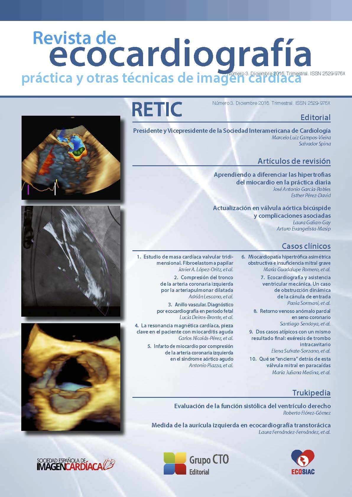
Three-dimensional valvular heart mass study. Papillary fibroelastoma
DOI:
https://doi.org/10.37615/retic.n3a4Keywords:
transesophageal echocardiography, 3D-echocardiography, aortic fibroelastoma.Abstract
This is a case of a 92 year-old lady who had a mass in the aortic valve leading to fibroelastoma supiction. 3D-transesophageal echo allowed to achieve a precise evaluation of the anatomy and structure of the mass and the valvular damage.
Downloads
Metrics
References
Jaffe W, Figured VM. An example of Lambl’s excresences by transesophageal echocardiogram: A commonly misinterpreted lesion. Echocardiography 2007; 24 (10): 1.086-1.089. DOI: https://doi.org/10.1111/j.1540-8175.2007.00533.x
Sydow K, Willems S, Reichenspurner H, Meinertz T. Papillary fibroelastomas of the heart. Thorac Cardiovasc Surg 2008; 56: 9. DOI: https://doi.org/10.1055/s-2007-989281
Braunwald E. Tumores que afectan al sistema cardiovascular. Cap. 85. En: Braunwald E (ed.). Tratado de cardiología. Texto de medicina cardiovascular. 10.ª ed. Elservier Barcelona (España). 2015; 1.869-1.870.
Bogaert J, Dymarkowski S, Taylor A, et al. Cardiac masses. Cap. 13. En: Bogaert J, Dymarkowski S, Taylor A, et al. Clinical Cardiac MRI. 2.nd ed. Berlin (Alemania): Springer; 2012; 418-432. DOI: https://doi.org/10.1007/978-3-642-23035-6
Herzog B, Greenwood J, Plein S. Cardiovascular Magnetic Resonance - Pocket Guide Cardiac Masses. 1.st ed. 2013; 102-107.
Saric M, Armour AC, Arnaout MS, et al. Guidelines for the use of echocardiography in the evaluation of the cardiac source of embolism. J Am Soc Echocardiogr 2016; 29 (1): 1-42. DOI: https://doi.org/10.1016/j.echo.2015.09.011
Buck T, Fanke A, Monaghan MJ (eds.). Cardiac tumors and sources of embolism. Cap. 11. En: Buck T, Fanke A, Monaghan MJ (eds.). Three-dimensional echocardiography. 2.nd ed. Springer (Alemania); 2014; 258-259.
Badano L, Lang R, Zamorano J (eds.). Three-dimensional echocardiography to asses intra-cardiac masses. Cap. 11. En: Badano L, Lang R, Zamorano J (eds.).Textbook of Real-time Three Dimensional Echocardiography. London; Springer; 2010.; 111-120. DOI: https://doi.org/10.1007/978-1-84996-495-1_11
Downloads
Published
How to Cite
Issue
Section
License
Copyright (c) 2016 Javier A. López-Opitz, Oscar Moreno-Urrutia, Jennifer Lara-Melo, Nilton Silva Durán

This work is licensed under a Creative Commons Attribution-NonCommercial-NoDerivatives 4.0 International License.
RETIC is distributed under the Creative Commons Attribution-NonCommercial-NoDerivatives 4.0 International (CC BY-NC-ND 4.0) license https://creativecommons.org/licenses/by-nc-nd/4.0 which allows sharing, copying and redistribution of the material in any medium or format, under the following terms:
- Attribution: you must give appropriate credit, provide a link to the license, and indicate if changes were made. You may do so in any reasonable manner, but not in any way that suggests that the licensor endorses you or your use.
- Non-commercial: you may not use the material for commercial purposes.
- No Derivatives: if you remix, transform or build upon the material, you may not distribute the modified material.
- No Additional Restrictions: you may not apply legal terms or technological measures that legally restrict others from doing anything permitted by the license.


