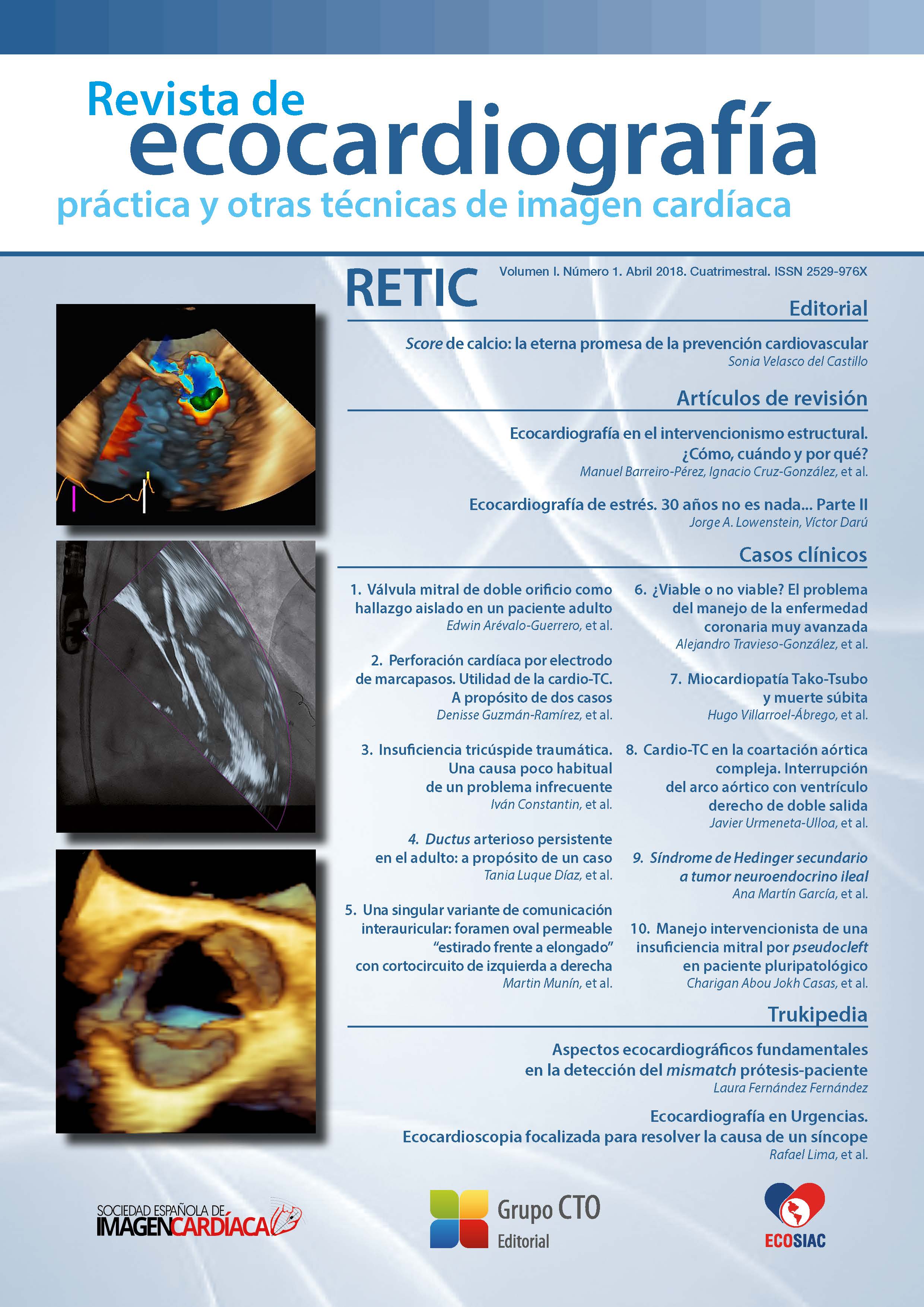
A unique variant of atrial septal defect: "stretched vs. elongated" patent foramen ovale with left-to-right shunting
DOI:
https://doi.org/10.37615/retic.v1n1a8Keywords:
stretched patent foramen ovale, atrial septal defect, septum primum, septum secundum.Abstract
We present the case of a 79 year old patient who attended the clinic due to heart failure. The patient had atrial fibrillation, mitral regurgitation, significant dilatation of the right chambers and preserved left ventricular ejec- tion fraction. The transesophageal echocardiography examination showed a significant separation between the septum primum and secundum, a finding compatible with a large “stretched” patent foramen ovale, with unidirectional continuous flow from left to right, behaving functionally as an atrial septal defect: “stretched” patent foramen ovale or “valve incompetent”.
Downloads
Metrics
References
Hagen PT, Scholz DG, Edwards WD. Incidence and size of patent foramen ovale during the first 10 decades of life: an autopsy study of 965 normal hearts. Mayo Clin Proc 1984; 59 (1): 17-20. DOI: https://doi.org/10.1016/S0025-6196(12)60336-X
Lechat P, Mas JL, Lascault G, et al. Prevalence of patent foramen ovale in patients with stroke. N Engl J Med 1988; 318 (18): 1.148-1.152. DOI: https://doi.org/10.1056/NEJM198805053181802
Ortega Trujillo JR, Suárez de Lezo Herreros de Tejada J, García Quintana A, et al. Transcatheter closure of patent foramen ovale in patients with platypnea-orthodeoxia. Rev Esp Cardiol 2006; 59 (1): 78-81. DOI: https://doi.org/10.1016/S1885-5857(06)60054-6
Calvert PA, Rana BS, Kydd AC, et al. Patent foramen ovale: anatomy, outcomes, and closure. Nat Rev Cardiol 2011; 8 (3): 148-160. DOI: https://doi.org/10.1038/nrcardio.2010.224
Wu CC, Chen WJ, Chen MF, et al. Left-to-right shunt through patent foramen ovale in adult patients with left-sided cardiac lesions: a transesophageal echocardiographic study. Am Heart J 1993; 125 (5 Pt 1): 1.369-1.374. DOI: https://doi.org/10.1016/0002-8703(93)91009-4
Ho SY, McCarthy KP, Rigby ML. Morphological features pertinent to interventional closure of patent oval foramen. J Interv Cardiol 2003; 16 (1): 33-38. DOI: https://doi.org/10.1046/j.1540-8183.2003.08000.x
Sakagianni K, Evrenoglou D, Mytas D, et al. Platypnea-orthodeoxia syndrome related to right hemidiaphragmatic elevation and a ‘stretched’ patent foramen ovale. BMJ Case Rep 2012; 10: 7.735. DOI: https://doi.org/10.1136/bcr-2012-007735
Downloads
Published
How to Cite
Issue
Section
License
Copyright (c) 2018 Martin Munín, Diego Xavier Chango Azanza, Noelia Pérez, Ignacio Raggio, Julieta Paolini

This work is licensed under a Creative Commons Attribution-NonCommercial-NoDerivatives 4.0 International License.
RETIC is distributed under the Creative Commons Attribution-NonCommercial-NoDerivatives 4.0 International (CC BY-NC-ND 4.0) license https://creativecommons.org/licenses/by-nc-nd/4.0 which allows sharing, copying and redistribution of the material in any medium or format, under the following terms:
- Attribution: you must give appropriate credit, provide a link to the license, and indicate if changes were made. You may do so in any reasonable manner, but not in any way that suggests that the licensor endorses you or your use.
- Non-commercial: you may not use the material for commercial purposes.
- No Derivatives: if you remix, transform or build upon the material, you may not distribute the modified material.
- No Additional Restrictions: you may not apply legal terms or technological measures that legally restrict others from doing anything permitted by the license.


