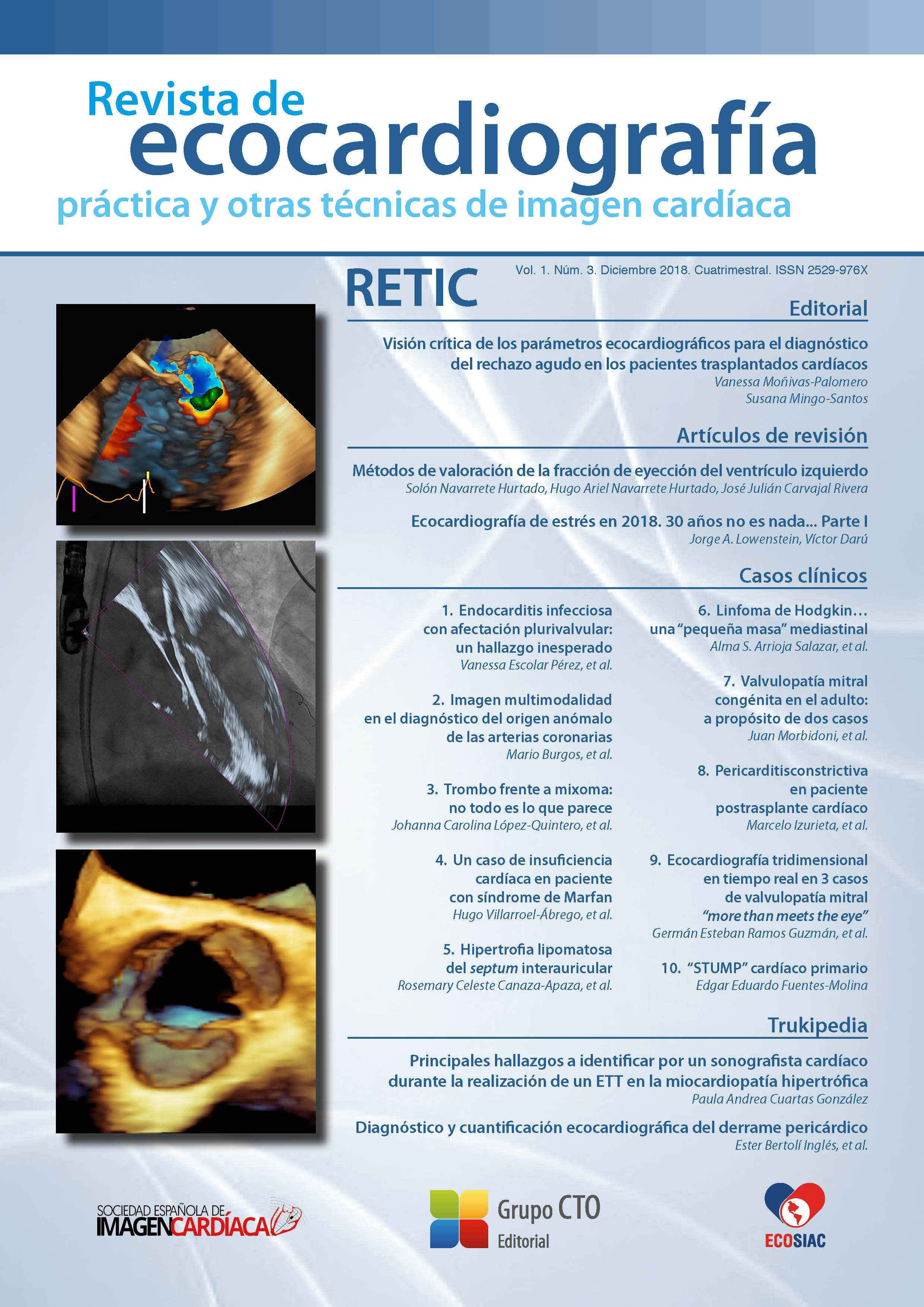
Lipomatous hypertrophy of the interatrial septum
DOI:
https://doi.org/10.37615/retic.v1n3a8Keywords:
lipomatous hypertrophy of the interatrial septum, three-dimensional echocardiography, two-dimensional echocardiography.Abstract
Lipomatous hypertrophy of the interatrial septum (HLSIA) is a benign and infrequent septal abnormality. We present the incidental finding of HLSIA diagnosed by transesophageal echocardiography (TEE) 2D/3D in an obese patient, of the third age, in a study performed evaluation of mitral regurgitation severity. The typical morphological characteristics (sign of the “hourglass”, septal thickness bigger than 20 mm) make easy the diagnosis. Although there is some controversy about its proper denomination due to its histopathological characteristics, HLSIA can be a cause of cardiac arrhythmias, superior vena cava syndrome, or be erroneously diagnosed as a malignant tumor. It is usually an incidental finding, with asymptomatic evolution and good prognosis.
Downloads
Metrics
References
Goldstein S. Normal Anatomic Variants and Artifacts. Lang R, Godstein A, Kronzon B. ASE’s Comprehensive Echocardiography, 2.ª Ed. Elsevier. Philadelphia, 2016; 642-643.
Augoustides J, Weiss S, Ochroch A, et al. Análisis del tabique interauricular mediante ecocardiografía transesofágica en pacientes adultos con cirugía cardíaca: variantes anatómicas y correlación con foramen oval permeable. J Cardiothorac Vasc Anesth. 2005; 19 (2): 146-149.
Chicago, Illin McAllister HA, Fenoglio J. Tumors of the cardiovascular system. Washington. Armed Forces Institute of Pathology, 1978.
Donnino L, Benenstein L, Freedberg S. Lipomatous Atrial Septal Hypertrophy: A Review of Its Anatomy, Pathophysiology, Multimodality Imaging, and Relevance to Percutaneous Interventions, o interatrial percutaneous. J Am Soc Echocardiogr. 2016; 29 (8): 717-723.
Reyes J VR. Lipomatous hypertrophy of the cardiac interatrial septum. A report of 38 cases and review of the literature. Am J Clin Pathol 1979; 72: 785.
Cabrera J, Zunen Y, Sarmientos Valiente J. Lipomatous hypertrophy of the interatrial septum: Myth or reality? Rev Fed Arg Cardiol 2011; 40 (3) 2.015.
Søholm H, Iversen K, Olsen PS, et al. Superior vena cava syndrome as a rare complication to lipomatous atrial septal hypertrophy. Eur Heart J Cardiovasc Imaging 2013; 14 (7): 717.
Heyer C, Kagel T, Lemburg S, et al. Lipomatous hypertrophy of the interatrial septum: a prospective study of incidence, imaging findings, and clinical symptoms. Cientific letters Chest 2003; 124: 2.068-2.073.
Downloads
Published
How to Cite
Issue
Section
License
Copyright (c) 2018 Rosemary Celeste Canaza-Apaza, Gustavo Restrepo-Molina, Edwin Arévalo-Guerrero, Jorge López, Karen Estupiñan

This work is licensed under a Creative Commons Attribution-NonCommercial-NoDerivatives 4.0 International License.
RETIC is distributed under the Creative Commons Attribution-NonCommercial-NoDerivatives 4.0 International (CC BY-NC-ND 4.0) license https://creativecommons.org/licenses/by-nc-nd/4.0 which allows sharing, copying and redistribution of the material in any medium or format, under the following terms:
- Attribution: you must give appropriate credit, provide a link to the license, and indicate if changes were made. You may do so in any reasonable manner, but not in any way that suggests that the licensor endorses you or your use.
- Non-commercial: you may not use the material for commercial purposes.
- No Derivatives: if you remix, transform or build upon the material, you may not distribute the modified material.
- No Additional Restrictions: you may not apply legal terms or technological measures that legally restrict others from doing anything permitted by the license.


