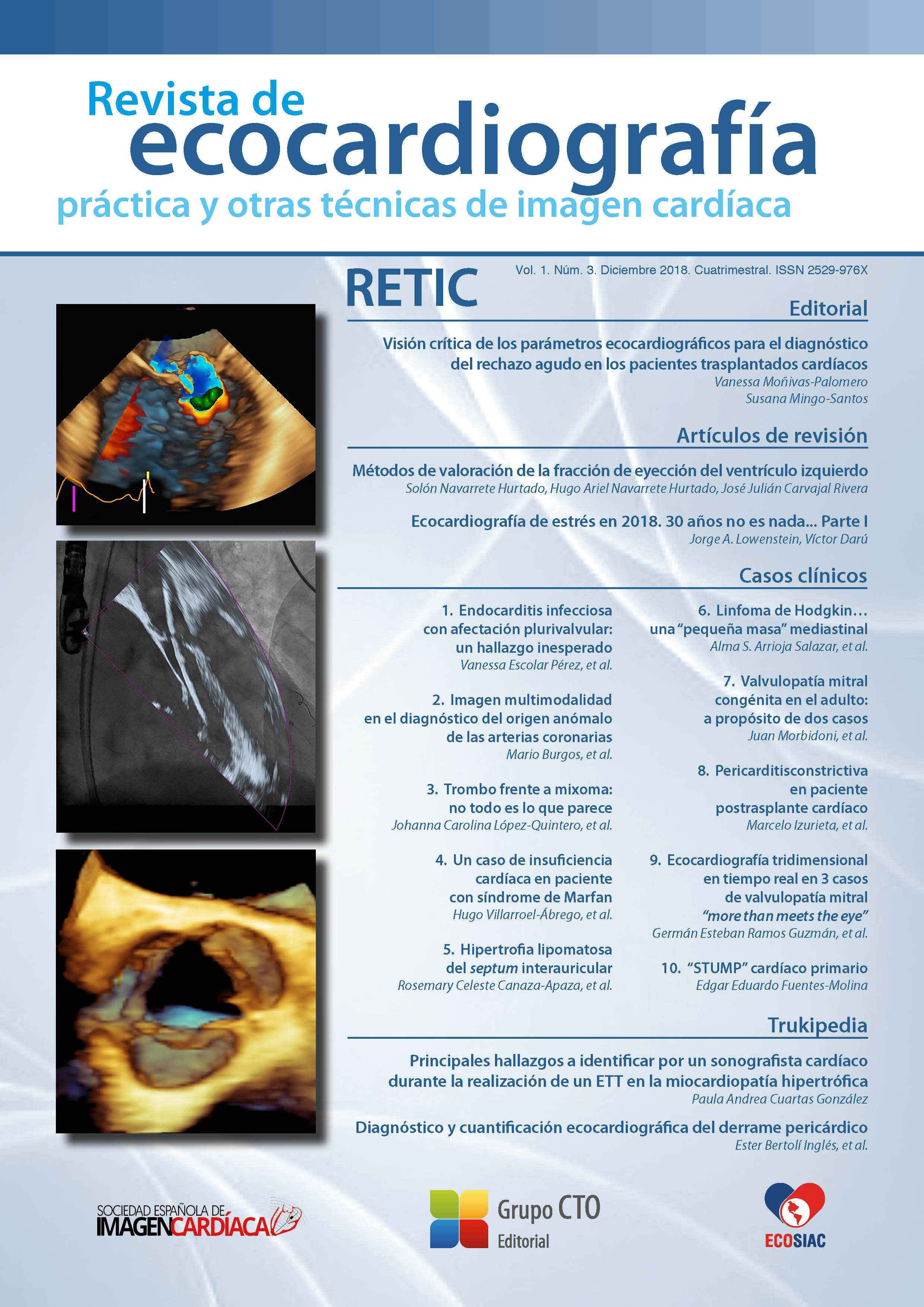
Ecocardiografía tridimensional en tiempo real en 3 casos de valvulopatía mitral “more than meets the eye”
DOI:
https://doi.org/10.37615/retic.v1n3a12Palabras clave:
ecocardiografía transesofágica tridimensional, trombosis anillo protésico mitral, prolapso valvular mitral, endocarditis perforada.Resumen
La ecocardiografía transesofágica tridimensional (ETE-3D) ha surgido en los últimos años como una herramienta de gran ayuda a la técnica bidimensional, en especial en lo que respecta al estudio de la válvula mitral por su localización en el campo cercano, lo que permite una evaluación exacta y detallada de la misma. Se presenta, a través de la descripción 3 casos (trombosis de anillo protésico, prolapso valvular y perforación por endocarditis), las ventajas que la imagen tridimensional en tiempo real puede ofrecer en la práctica diaria.
Descargas
Métricas
Citas
Laplace G, Lafitte S, Labèque J, et al. Clinical significance of early thrombosis after prosthetic mitral valve replacement: a postoperative monocentric study of 680 patients. J Am Coll Cardiol 2004; 43: 1.283-1.290.
Dangas G, Weitz J, Giustino G, et al. Prosthetic Heart Valve Thrombosis. J Am Coll Cardiol 2016; 68: 2.670-2.689.
Ozkan M, Gürsoy OM, Astarcıoğlu MA, et al. Real-time three dimensional transesophageal echocardiography in the assessment of mechanical prosthetic mitral valve ring thrombosis. Am J Cardiol 2013; 1; 112 (7): 977-983.
Gürsoy OM, Karakoyun S, Kalçık M, Özkan M. The incremental value of RT three-dimensional TEE in the evaluation of prosthetic mitral valvering thrombosis complicated with thromboembolism. Echocardiography 2013; 30 (7): E198-201.
Benenstein R, Saric M. Mitral valve prolapse: role of 3D echocardiography in diagnosis. Curr Opin Cardiol 2012, 27: 465-476.
Addetia K, Mor-Avi V, Weinert L, et al. A New Definition for an Old Entity: Improved Definition of Mitral Valve Prolapse Using Three-Dimensional Echocardiography and Color-Coded Parametric Models. J Am Soc Echocardiogr 2014; 27: 8-16.
Faletra F, Demertzis S, Pedrazzini G, et al. Three dimensional transesophageal echocardiography in degenerative mitral regurgitation. J Am Soc Echocardiogr 2015; 28 (4): 437-448.
De Groot-de Laat LE, Ren B, McGhie J, Oei FB, et al. The role of experience in echocardiographic identification of location and extent of mitral valve prolapse with 2D and 3D echocardiography. Int J Cardiovasc Imaging 2016; 32 (8): 1.171-1.177.
Habib G, Hoen B, Tornos P, et al. Guidelines on the prevention, diagnosis, and treatment of infective endocarditis (new version 2009). European Heart Journal 2009; 30 (19): 2.369-2.413.
Salcedo EE, Quaife RA, Seres T, Carroll JD. A framework for systematic characterization of the mitral valve by realtime three-dimensional transesophageal echocardiography. J Am Soc Echocardiogr 2009; 22: 1.087-1.099.
Bhave NM, Addetia K, Spencer KT, et al. Localizing mitral valve perforations with 3D transesophageal echocardiography. JACC Cardiovascular Imaging 2013; 6 (3): 407.
Thompson KA, Shiota T, Tolstrup K, et al. Utility of three-dimensional transesophageal echocardiography in the diagnosis of valvular perforations. American Journal of Cardiology 2011; 107 (1): 100-102.
Descargas
Publicado
Cómo citar
Número
Sección
Licencia
Derechos de autor 2018 Germán Esteban Ramos Guzmán , Manuel Rodríguez Venegas , Mario Zapata Muñoz

Esta obra está bajo una licencia internacional Creative Commons Atribución-NoComercial-SinDerivadas 4.0.
RETIC se distribuye bajo la licencia Creative Commons Reconocimiento-NoComercial-SinDerivadas 4.0 Internacional (CC BY-NC-ND 4.0) https://creativecommons.org/licenses/by-nc-nd/4.0 que permite compartir, copiar y redistribuir el material en cualquier medio o formato, bajo los siguientes términos:
- Reconocimiento: debe otorgar el crédito correspondiente, proporcionar un enlace a la licencia e indicar si se realizaron cambios. Puede hacerlo de cualquier manera razonable, pero no de ninguna manera que sugiera que el licenciante lo respalda a usted o su uso.
- No comercial: no puede utilizar el material con fines comerciales.
- No Derivados: si remezcla, transforma o construye sobre el material, no puede distribuir el material modificado.
- Sin restricciones adicionales: no puede aplicar términos legales o medidas tecnológicas que restrinjan legalmente a otros de hacer cualquier cosa que permita la licencia.


