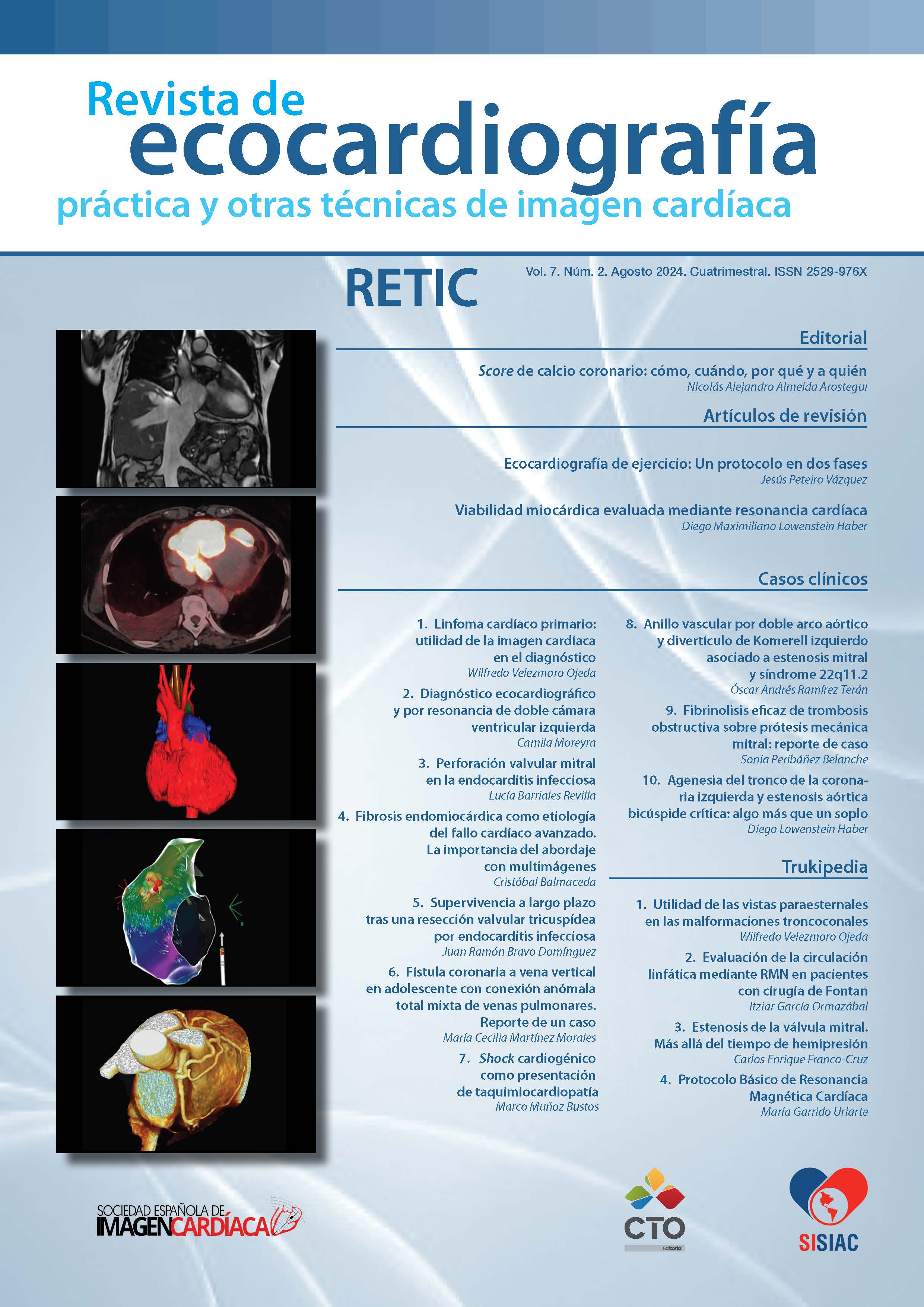
Viabilidad miocárdica evaluada mediante resonancia cardíaca
DOI:
https://doi.org/10.37615/retic.v7n2a3Palabras clave:
resonancia magnética cardíaca, viabilidad miocárdica, realce tardío con gadolinio, enfermedad coronaria, disfunción ventricular izquierdaResumen
La resonancia magnética cardíaca (RMC) con realce tardío de gadolinio es una técnica avanzada para evaluar la viabilidad miocárdica, esencial para la toma de decisiones sobre la revascularización en pacientes con enfermedad coronaria y disfunción ventricular izquierda. La RMC proporciona imágenes de alta resolución sin radiación ionizante, lo que la hace segura y repetible. Este artículo revisa la evolución de las técnicas de imagen para la evaluación de la viabilidad miocárdica, centrándose en la RMC y su capacidad para identificar el tejido miocárdico viable. Se discuten los fundamentos de la RMC, la importancia del gadolinio como agente de contraste y los criterios de transmuralidad de la fibrosis para determinar la viabilidad miocárdica. Además, se destacan estudios clave que han demostrado la precisión y relevancia clínica de la RMC en este contexto.
Descargas
Métricas
Citas
Allman KC, Shaw LJ, Hachamovitch R, Udelson JE. Myocardial viability testing and impact of revascularization on prognosis in patients with coronary artery disease and left ventricular dysfunction: a meta-analysis. JAm Coll Cardiol. 2002;39(7):1151-1158. doi: https://doi.org/10.1016/s0735-1097(02)01726-6 DOI: https://doi.org/10.1016/S0735-1097(02)01726-6
Strauss HW, Pitt B. Thallium-201 as a myocardial imaging agent. Circulation. 1972;46(4):647-650). doi: https://doi.org/10.1016/s0001-2998(77)80007-x DOI: https://doi.org/10.1016/S0001-2998(77)80007-X
Gould KL, Goldstein RA, Mullani NA, et al. Noninvasive assessment of coronary stenoses by myocardial perfusion imaging during pharmacologic coronary vasodilation. VIII. Clinical feasibility of positron cardiac imaging without a cyclotron using generator-produced rubidium-82. JAm Coll Cardiol. 1986;7(4):775-789). doi: https://doi.org/10.1016/s0735-1097(86)80336-9 DOI: https://doi.org/10.1016/S0735-1097(86)80336-9
Kim RJ, Wu E, Rafael A, et al. The use of contrast-enhanced magnetic resonance imaging to identify reversible myocardial dysfunction. N Engl J Med. 1999;341(8):489-497. doi: https://doi.org/10.1056/NEJM200011163432003
Wagner, A., Mahrholdt, H., Holly, T. A., Elliott, M. D., Regenfus, M., Parker, M., Klocke, F. J., Bonow, R. O., Kim, R. J., & Judd, R. M. (2003). Contrast-enhanced MRI and routine single photon emission computed tomography (SPECT) perfusion imaging for detection of subendocardial myocardial infarcts: an imaging study. The Lancet, 361(9355), 374-379. doi: https://doi.org/10.1016/S0140-6736(03)12389-6 DOI: https://doi.org/10.1016/S0140-6736(03)12389-6
Choi KM, Kim RJ, Gubernikoff G, et al. Transmural Extent of Acute Myocardial Infarction Predicts Long-Term Improvement in Contractile Function. Circulation. 2001;104:1101-1107 doi: https://doi.org/10.1161/hc3501.096798 DOI: https://doi.org/10.1161/hc3501.096798
Baer, FM, Theissen, P, Schneider, CA, Voth, E, Sechtem, U, Schicha, H, Erdmann, E. Dobutamine magnetic resonance imaging predicts contractile recovery of chronically dysfunctional myocardium after successful revascularization. J Am Coll Cardiol. 1998;31:1040–1048. doi:
1016/s0735-1097(98)00032-1). doi: https://doi.org/10.1016/s0735-1097(98)00032-1 DOI: https://doi.org/10.1016/S0735-1097(98)00032-1
Selvanayagam JB, Kardos A, Nicolson D, et al. Value of Delayed-Enhancement Cardiovascular Magnetic Resonance Imaging in Predicting Myocardial Viability After Surgical Revascularization. Circulation. 2004;110:1535-1541). doi: https://doi.org/10.1161/01.CIR.0000142045.22628.74 DOI: https://doi.org/10.1161/01.CIR.0000142045.22628.74
Kim, R. J., Wu, E., Rafael, A., Chen, E. L., Parker, M. A., Simonetti, O., ... & Judd, R. M. (2000). The use of contrast-enhanced magnetic resonance imaging to identify reversible myocardial dysfunction. New England Journal of Medicine, 343(20), 1445-1453. doi: https://doi.org/10.1056/NEJM200011163432003 DOI: https://doi.org/10.1056/NEJM200011163432003
Kwong, R. Y., Chan, A. K., Brown, K. A., Chan, C. W., Reynolds, H. G., Tsang, S., & Davis, R. B. (2006). Impact of unrecognized myocardial scar detected by cardiac magnetic resonance imaging on event-free survival in patients presenting with signs or symptoms of coronary artery disease. Circulation, 113(23), 2733-2743. doi: https://doi.org/10.1161/CIRCULATIONAHA.105.570648 DOI: https://doi.org/10.1161/CIRCULATIONAHA.105.570648
Velazquez EJ, Lee KL, Jones RH, et al. Coronary-artery bypass surgery in patients with left ventricular dysfunction. N Engl J Med. 2011;364(17):1607-1616. doi: https://doi.org/10.1056/NEJMoa1100356 DOI: https://doi.org/10.1056/NEJMoa1100356
Cleland J.G.F., Calvert M., Freemantle N., Arrow Y., Ball S.G., Bonser R.S., Chattopadhyay S., Norell M.S., Pennell D.J., Senior R. The Heart Failure Revascularisation Trial (HEART) Eur. J. Heart Fail. 2011;13:227–233. doi: https://doi.org/10.1093/eurjhf/hfq230 DOI: https://doi.org/10.1093/eurjhf/hfq230
Rahimi K, et al. Percutaneous coronary intervention in patients with severe ischaemic left ventricular dysfunction (REVIVED-BCIS2): an open-label, randomised controlled trial. Lancet. 2022;400(10360):760-768. doi: https://doi.org/10.1016/j.jchf.2018.01.024 DOI: https://doi.org/10.1016/j.jchf.2018.01.024
Wellnhofer E, Olariu A, Klein C, Gräfe M, Wahl A, Fleck E, Nagel E. Magnetic resonance low-dose dobutamine test is superior to SCAR quantification for the prediction of functional recovery. Circulation. 2004;109:2172-2174. doi: https://doi.org/10.1161/01.CIR.0000128862.34201.74 DOI: https://doi.org/10.1161/01.CIR.0000128862.34201.74
Publicado
Cómo citar
Número
Sección
Licencia
Derechos de autor 2024 Alvarenga Andrea, Cardozo Leandro, Escalante Exequiel, Imaz, Geronimo, Lowenstein Haber, Diego, Miguel Ángel Freis

Esta obra está bajo una licencia internacional Creative Commons Atribución-NoComercial-SinDerivadas 4.0.
RETIC se distribuye bajo la licencia Creative Commons Reconocimiento-NoComercial-SinDerivadas 4.0 Internacional (CC BY-NC-ND 4.0) https://creativecommons.org/licenses/by-nc-nd/4.0 que permite compartir, copiar y redistribuir el material en cualquier medio o formato, bajo los siguientes términos:
- Reconocimiento: debe otorgar el crédito correspondiente, proporcionar un enlace a la licencia e indicar si se realizaron cambios. Puede hacerlo de cualquier manera razonable, pero no de ninguna manera que sugiera que el licenciante lo respalda a usted o su uso.
- No comercial: no puede utilizar el material con fines comerciales.
- No Derivados: si remezcla, transforma o construye sobre el material, no puede distribuir el material modificado.
- Sin restricciones adicionales: no puede aplicar términos legales o medidas tecnológicas que restrinjan legalmente a otros de hacer cualquier cosa que permita la licencia.


